


Written by: Liz Paton, MSc
Brain Anatomy Overview
The human brain is the most complex part of our body and the organ that defines our humanity. The brain is composed of three major sections: The cerebrum, cerebellum, and brainstem.
The cerebrum is the uppermost and largest part of the brain. It is split by a central fissure into the right and left hemispheres and is also divided into four major lobes. The cerebrum controls several functions such as learning, emotions and movement, to name a few.
The cerebellum is situated underneath the cerebrum and is used to coordinate our movements, posture and balance. The cerebellum is found below the temporal and occipital lobes, and it is the largest structure of our hindbrain. The cerebellum is broken up into three lobes: the flocculonodular lobe, anterior lobe and posterior lobe.
The brainstem connects the brain to and is structurally continuous with the spinal cord. It is responsible for involuntary functions such as breathing, heart rate, sleep cycles, vomiting, digestion, coughing and sneezing and several other autonomic functions. The brainstem consists of the medulla oblongata, pons, and midbrain.
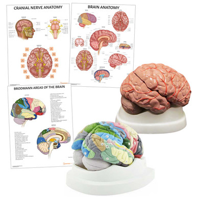
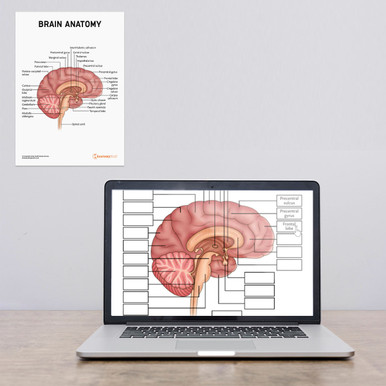
Our Brain Anatomy Chart visualises this perfectly.
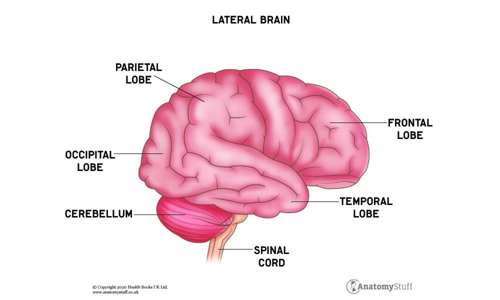
Lobes of the Brain
Frontal lobe
The frontal lobe is the largest lobe of the brain, located directly behind the forehead. It is responsible for your self-awareness, emotions, planning, judgement, movement, and speech. It is involved with memory, impulse control, and social behaviour. The frontal lobe is separated from the parietal lobe by the central sulcus and separated from the temporal lobes by the lateral sulcus.
Parietal lobe
The parietal lobe is located above the temporal lobe and between the frontal lobe and occipital lobe. Containing the sensory cortex, it is responsible for the sense of touch, pain and temperature. It is also responsible for interpreting language, hearing, memory, and spatial and visual perception.
Temporal lobe
The temporal lobes are the second-largest lobes of the brain, located behind the ears. They are involved with memory, hearing, organisation and understanding language.
Occipital lobe
The occipital lobe is the smallest lobe of the brain, located posterior to the parietal and temporal lobes, lying beneath the skull’s occipital bone. It is responsible for interpreting vision.
Blood Supply of the Brain
The main blood supply to the brain comes from the carotid arteries and the vertebral arteries. The carotid arteries branch off from the aorta (the largest artery in your body found by the heart) and are broken into the internal and external carotid artery. The internal carotid artery and the vertebral arteries come together to form an anastomosis, a complex network of blood vessels that provide an alternative route for blood to move through if an injury to the brain occurs. This particular anastomosis is called the circle of Willis.
Circle of Willis
The circle of Willis is found on the inferior (bottom) side of the brain and is responsible for supplying oxygenated blood to more than 80% of the cerebrum. It is composed of the anterior cerebral arteries, anterior communicating artery, internal carotid arteries, posterior cerebral arteries, and posterior communicating arteries.
Cranial Nerves
The brain is the central organ of our nervous system, consisting of twelve pairs of cranial nerves. The twelve cranial nerves are as follows:
I. Olfactory nerve
The olfactory nerve is the first and shortest of the cranial nerves. It conveys sensory information related to your sense of smell.
II. Optic nerve
The optic nerve conveys sensory information to the brain regarding your sense of vision.
III. Oculomotor nerve
The oculomotor nerve helps control the muscle movements of your eyes. It also constricts the pupil exposed to bright lighting and helps your lens adjust when looking at near or far objects.
IV. Trochlear nerve
The trochlear nerve is also involved with the movements of your eyes by supplying the superior oblique muscle. It allows your eye to point downward and inward.
V. Trigeminal nerve
The trigeminal nerve is the largest and most complex of the cranial nerves and is involved with facial sensation. It is divided into three parts. The ophthalmic part provides sensation to parts of your eyes, nose, eyelids and forehead. The maxillary part provides sensation to the middle of your face, part of your nose, upper teeth and lower eyelids. The mandibular part gives sensation to the lower part of your face, including the tongue and lower teeth. A significant action fired by the trigeminal nerve is allowing you to masticate (chew) your food.
VI. Abducens nerve
The abducens nerve is yet another nerve that is involved with the movements of your eyes by supplying the lateral rectus muscle. It allows you to look outwards.
VII. Facial Nerve
The facial nerve is the nerve involved with your facial expression, sense of taste and sensation of your ear. It comprises three different nuclei: the motor nucleus, the parasympathetic nuclei and the sensory nucleus.
VIII. Vestibulocochlear nerve
The vestibulocochlear nerve is involved with your balance and hearing. It branches off into two different components, the vestibular nerve and the cochlear nerve. The vestibular nerve helps you maintain balance, while the cochlear nerve helps you hear.
IX. Glossopharyngeal nerve
The glossopharyngeal nerve allows you to swallow and controls your gag reflex and the taste sensation on the back of your tongue.
X. Vagus nerve
The vagus nerve is involved with motor, sensory and parasympathetic functions. The motor part of the vagus nerve gives off movements in your throat responsible for the initiation of swallowing. The sensory part of the vagus nerve gives you sensation on the outer part of your ear, in your throat, your abdominal organs and heart, and contributes to your sensation of taste. The parasympathetic part of the vagus nerve regulates your heart rhythm and innervates your lungs and gastrointestinal tract.
XI. Accessory nerve
The accessory nerve innervates the sternoclediomastoid and trapezius muscles and allows you to rotate, extend and flex your neck and shoulders.
XII. Hypoglossal nerve
Finally, the hypoglossal nerve supplies the muscles of your tongue.
Exercising the Brain
Exercising the brain is important to improve your cognitive ability (memory and daily functionality), especially as you get older. A few brain exercises include: completing jigsaw puzzles, learning new skills such as a new language, playing music, and reading.
Free Download PDFs
View All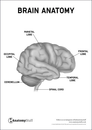
Related Products
View All
save
30%
AnatomyStuff
Brain Anatomy Poster / Worksheet (Interactive & Printable PDF)
Now
£3.50
Inc VAT
Was
£5.00
Inc VAT


