Written by: Liz Paton, MSc
Knee Anatomy Overview
The knee helps to provide stability and also helps with shock absorption when weight-bearing. This strong joint helps to support the weight of our upper body. The knee allows essential movement when carrying out various activities such as walking, running, sitting and standing.
Take a look at our Knee Anatomy Revision Worksheets for some extra help on learning this topic.
The Motion of the Knee
The knee is a modified hinge synovial joint. The reason why the knee is a modified joint is that as well as allowing flexion and extension, the knee also provides a small amount of medial and lateral rotation.
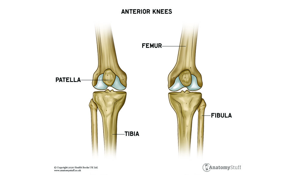
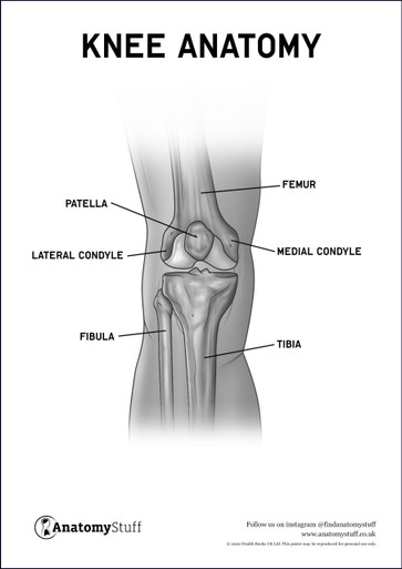
Bones of the Knee
The knee is made up of the femur (the bone in the thigh, the tibia (one of the bones of the lower leg, and the patella (the knee cap). There are two articulations which consist of the tibiofemoral joint where the tibia meets the femur and the patellofemoral joint, where the patella meets the femur. The knee joint is surrounded by a joint capsule for protection and lubrication when bending and to increase strength.
The Cartilage of the Knee
As well as bones, the joint is also made up of cartilage. The cartilage in the knee is called the meniscus which is made of a tough rubber-like material that helps with shock absorption. There is a lateral and medial meniscus in each knee which are located in between the tibia and femur. The menisci help to increase the stability of the joint and prevents bones from coming into contact during various movements.
Muscles of the Knee
The muscles and tendons move the knee joint via a system where the muscles contract to pull the tendons which are attached to the bone. The muscles of the knee consist of the hamstrings, the quadriceps and the muscles in the lower leg.
The quadriceps femoris muscles are formed by four muscles situated in the front section of the thigh. These muscles include vastus lateralis, vastus medialis, vastus intermedius, and rectus femoris and they extend the knee joint.
The three muscles in the hamstring section which is situated in the back of the thigh flex the knee joint. The muscles in this group are biceps femoris, semimembranosus and semitendinosus. Another muscle that assists with flexion of the knee is the gastrocnemius muscle which is situated at the back of the lower leg (calf).
As mentioned previously, the knee allows for medial and lateral rotation. The popliteus muscle is situated at the back of the knee which alongside two muscles in the hamstring group (semimembranosus and semitendinosus) allows for medial rotation of the knee. The other muscle from this group is the biceps femoris which provides lateral rotation of the knee.
Ligaments of the Knee
The ligaments in the knee joint provide stability to the knee during different movements. These ligaments are crucial to prevent our knees from dislocating.
The patella ligament is a thick band of fibrous tissue which connects the patella to the tibia which helps to stabilise the patella.
There are two collateral ligaments that sit on either side of the knee called the medial and lateral collateral ligaments. These ligaments help to stabilise the knee by preventing too much sideways motion and reducing rotational forces applied to the knee. The patella ligament and collateral ligaments are known as extracapsular ligaments because they are located outside the knee joint.
The cruciate ligaments are known as intracapsular because they are located inside the knee joint. These ligaments control the forwards and backwards motion of the knee. These are the anterior and posterior cruciate ligament and together they create a “cross-shape”. The anterior cruciate ligaments help to prevent the tibia from moving backwards relative to the femur and also prevents the knee from excessive extension. The posterior cruciate ligament prevents anterior movement of the tibia relative to the femur and prevents the knee from excessive flexion.
Blood Vessels of the Knee
The blood supply to the knee contains an intricate network of blood vessels; this is known as genicular anastomosis. The reason for this is so that in the event of an injury to the knee, the blood supply can find another route in case some of the blood supply becomes damaged.
The superior medial genicular artery, inferior medial genicular artery, superior lateral genicular artery, inferior lateral genicular artery and the middle genicular artery all arise from the popliteal artery.
Blood supply to the popliteal artery is provided by the descending genicular artery which stems from the femoral artery.
Nerves of the Knee
The femoral nerve supplies innervation to the knee joint. The knee joint also received innervation from the tibial and common peroneal nerves as well as a branch from the obturator nerve.