


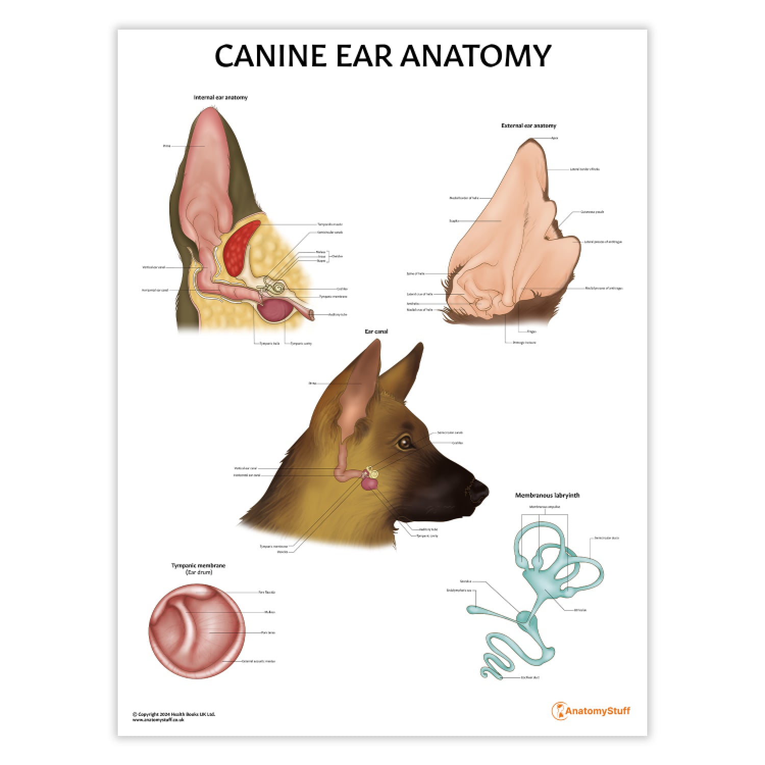

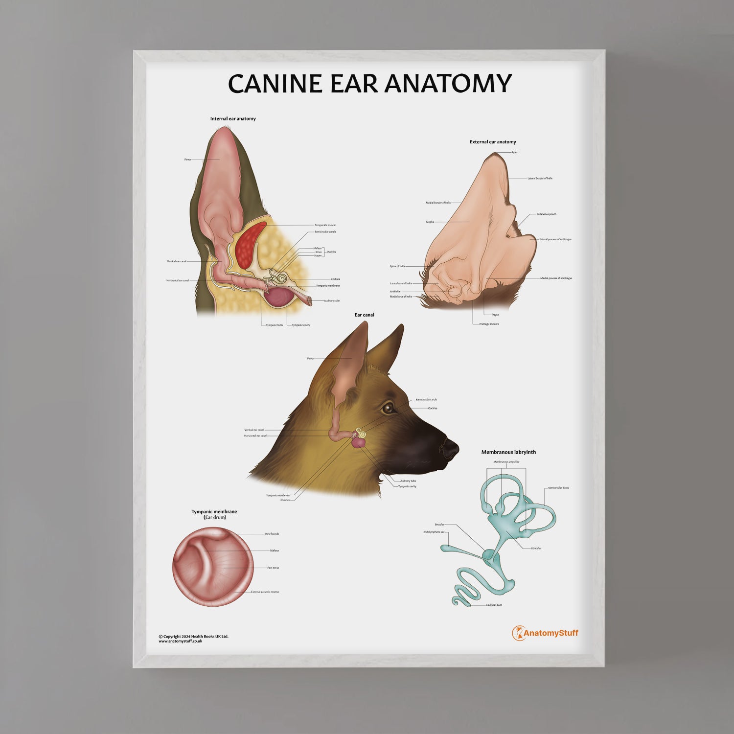

The Canine Ear Anatomy Laminated Chart/Poster is a valuable tool for veterinary professionals or students. This precise and anatomically accurate chart showcases detailed views of the outer and inner canine ear in stunning clarity. Perfect for educational purposes at the undergraduate level or as a visual aid in clinical settings.
Designed by a professional medical illustrator and exclusive to AnatomyStuff, our Canine Ear Anatomy Chart showcases the following in great anatomical details:
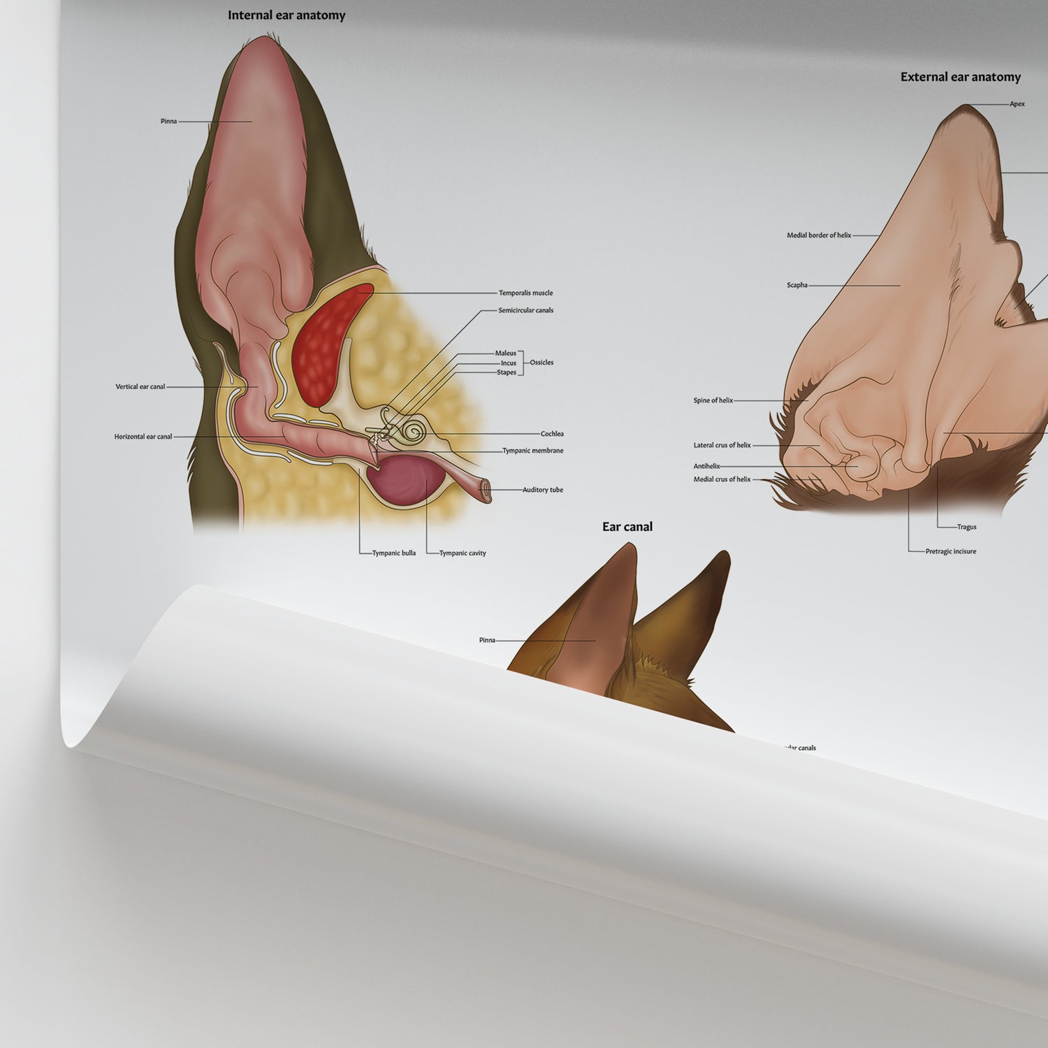
- Large cross-section view of the inner canine ear
- Anterior view of the membranous labyrinth
- Anterior view of the tympanic membrane
- Medial view of the ear canal
- External view of the external canine ear
Our collection offers a range of display options to meet your needs:
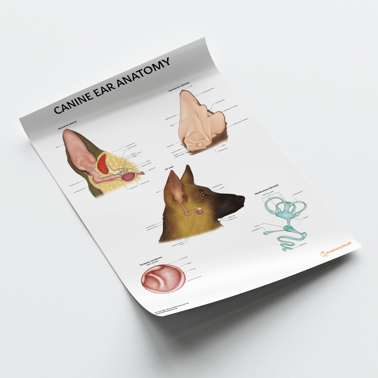
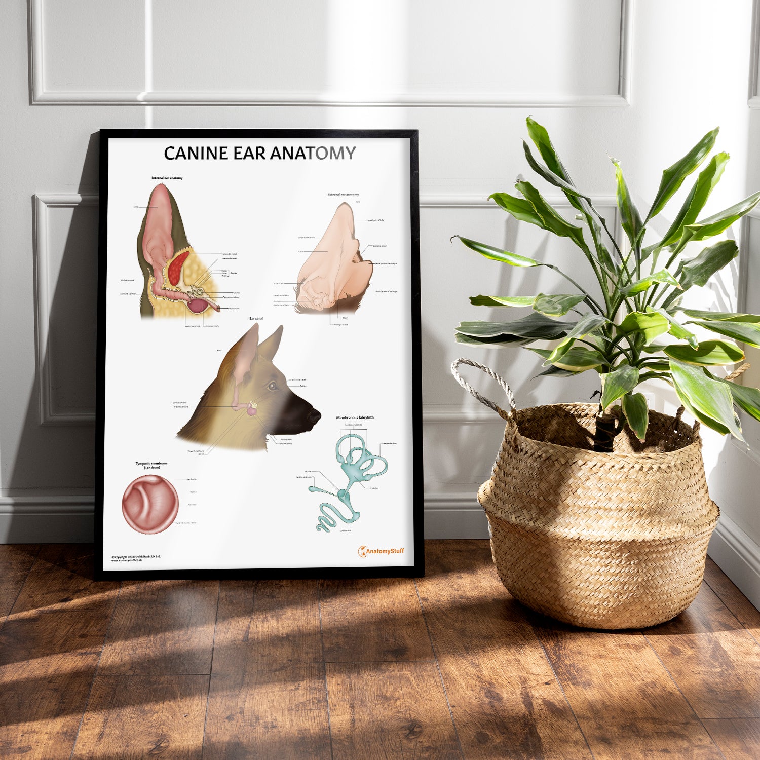
- Laminated Poster (50 x 65 cm): Ideal for clinic displays, featuring clearly labelled anatomical details. The durable lamination allows for annotation with washable markers (not supplied).
- Classic Semi-Glossy Prints: 45 x 60 cm, 60 x 80 cm, and 70 x 100 cm sizes. The semi-glossy finish enhances colours with a subtle shine, adding vibrancy to any setting.
- Framed Prints: Offered in black or white frames and sizes 45 x 60 cm, 60 x 80 cm, and 70 x 100 cm. Features 170 gsm matte paper with a smooth, non-reflective finish. Ready-to-hang with a durable pine wood frame.
