


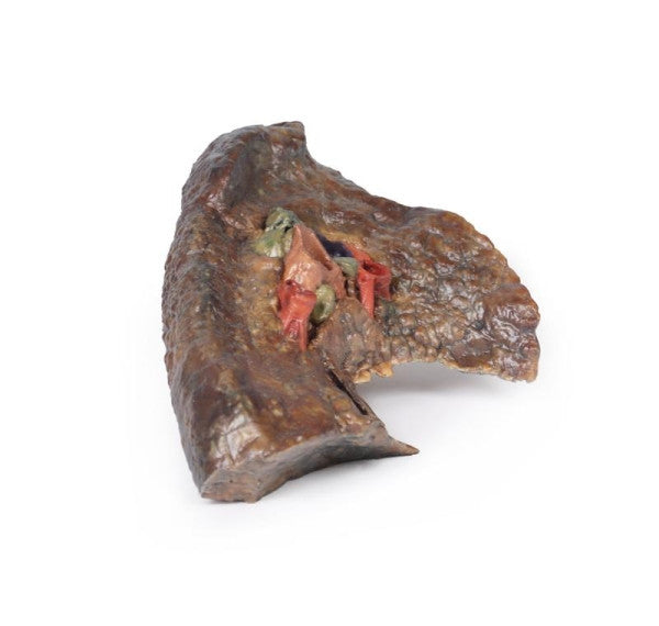
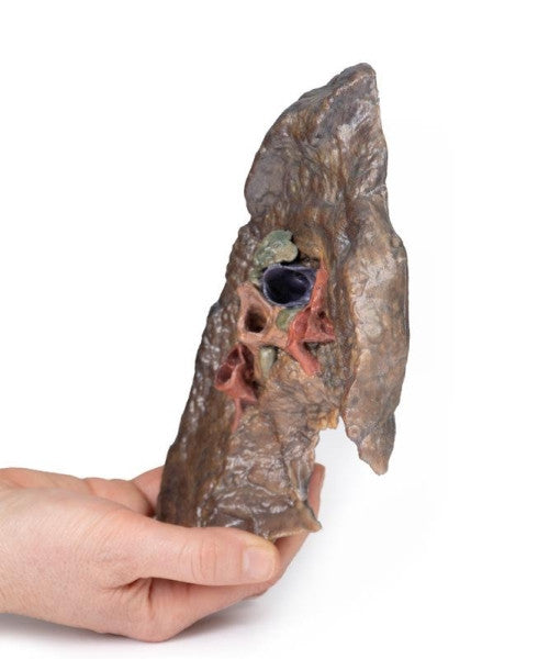
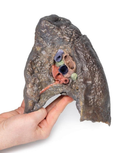
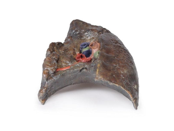

Hilum of the Left and Right Lungs
The lung's hilum connects to the body via the pulmonary ligament, housing vital components like the pulmonary arteries, main bronchi, and pulmonary veins. A sagittal cut reveals crucial structures, including the aortic arch impression, bronchi divisions, pulmonary vessels, and lymph nodes, offering detailed anatomical insight.
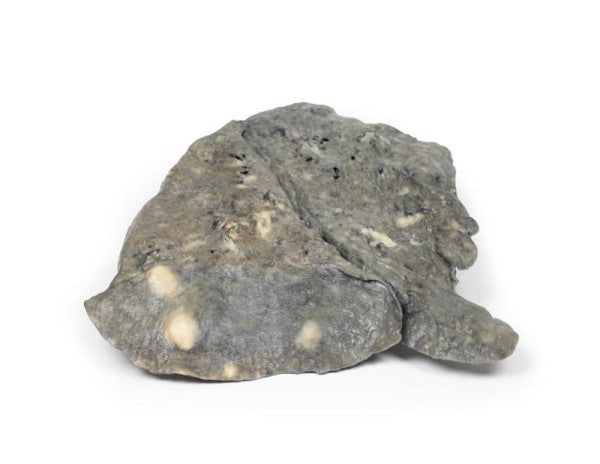
Lung (Staphylococcus Aureus Abscesses)
The bisected right lung exhibits multiple irregular abscess cavities, including a 4x3 cm cavity in the lower lobe's apex. Another 3x2 cm abscess in the upper lobe's apex is surrounded by consolidation. Small abscesses and pus-obstructed bronchi indicate Staphylococcal lung abscesses in an immunosuppressed patient, confirmed by Staph. aureus cultures.
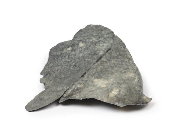
Lung (Cystic Fibrosis)
Bronchial changes, notably bronchiectasis, are widespread, with dilated bronchi and purulent material. The upper lobe apex shows a "honeycomb" change, and multiple abscesses appear in the lower lobe. Severe fibrosis and consolidation in the lower lobe's base are indicative of cystic fibrosis.
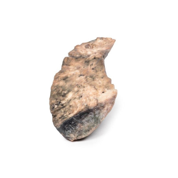
Lung (Multiple Secondary Carcinoma Deposits in the Lung and Pleura)
The intact left lung exhibits scattered pale tumour nodules, some confluently near the hilum. Hilar lymph nodes and small nodules beneath thickened pleura on various surfaces show metastatic adenocarcinoma deposits. A post-mortem revealed ovarian adenocarcinoma with metastases in multiple organs.
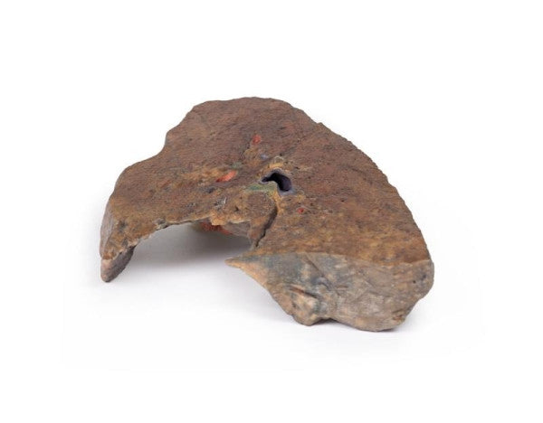
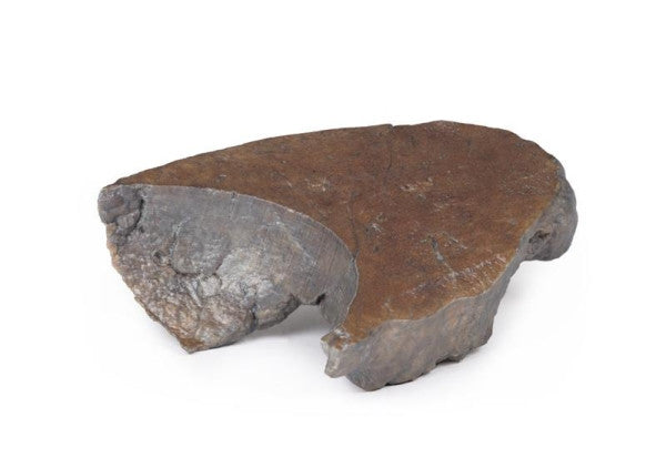
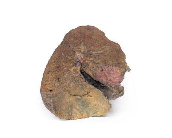
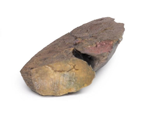
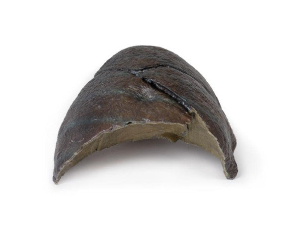
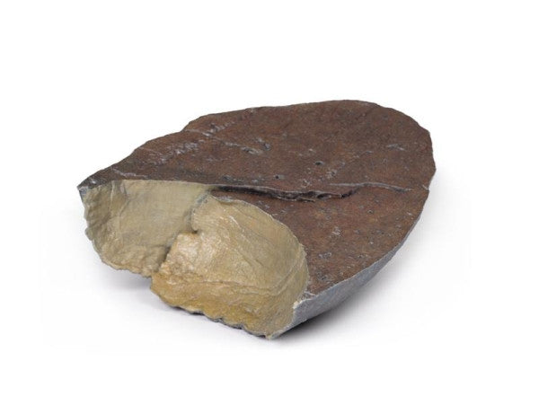
Lung Slab (Hilum Removed)
This parasagittal-dissected lung reveals the cardiac impression by the left ventricle and a concave diaphragmatic surface above the diaphragm. The bronchi's subdivision level remains unclear due to lateral dissection and pleura details are absent, but a diaphragmatic recess typically exists between the diaphragmatic impression and the diaphragm.
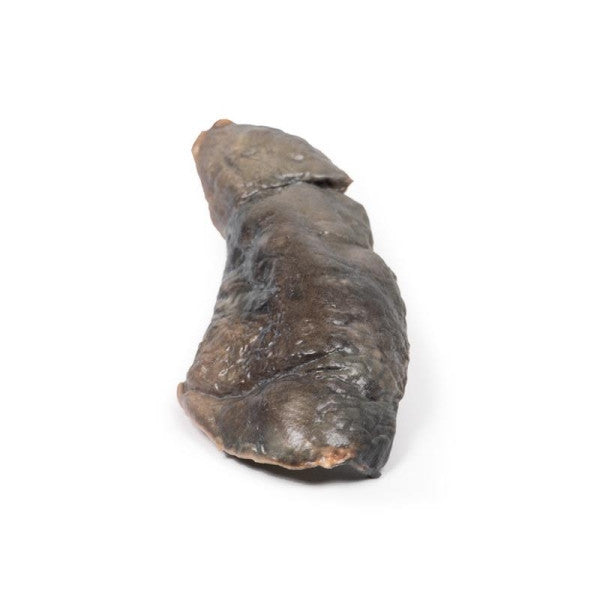
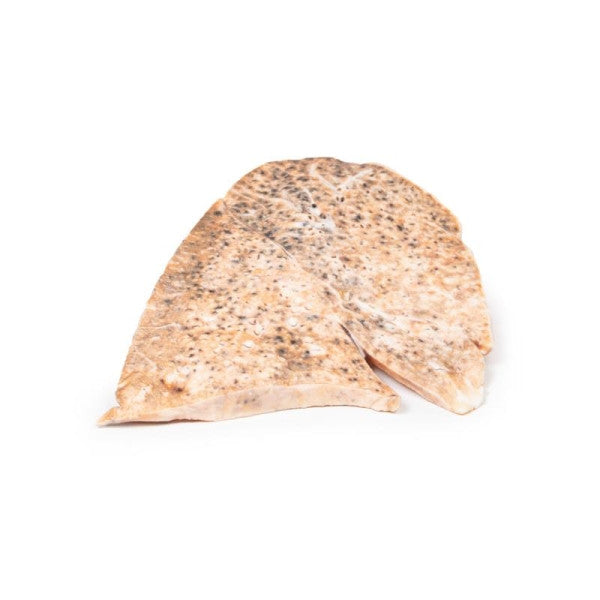
Right Lung (Miliary Tuberculosis)
The right lung, longitudinally sliced, exhibits mildly ecstatic bronchi and numerous small pale yellow nodules, each less than 1 mm in diameter. These tubercles, resembling millet seeds, indicate miliary tuberculosis.
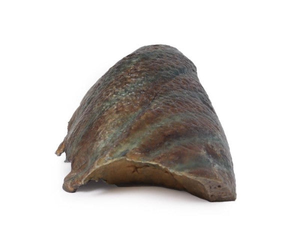
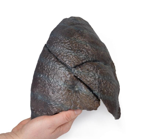
Right Lung (Hilum Removed)
This 3D model provides a detailed view from apex to base, it highlights the well-defined lobes, oblique, and horizontal fissures, and rib impressions. The concave diaphragmatic surface reflects the natural doming caused by the liver beneath in vivo.