


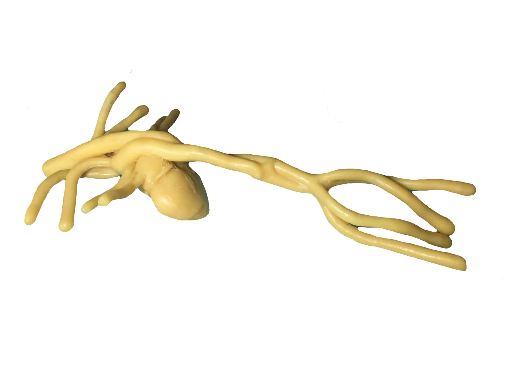
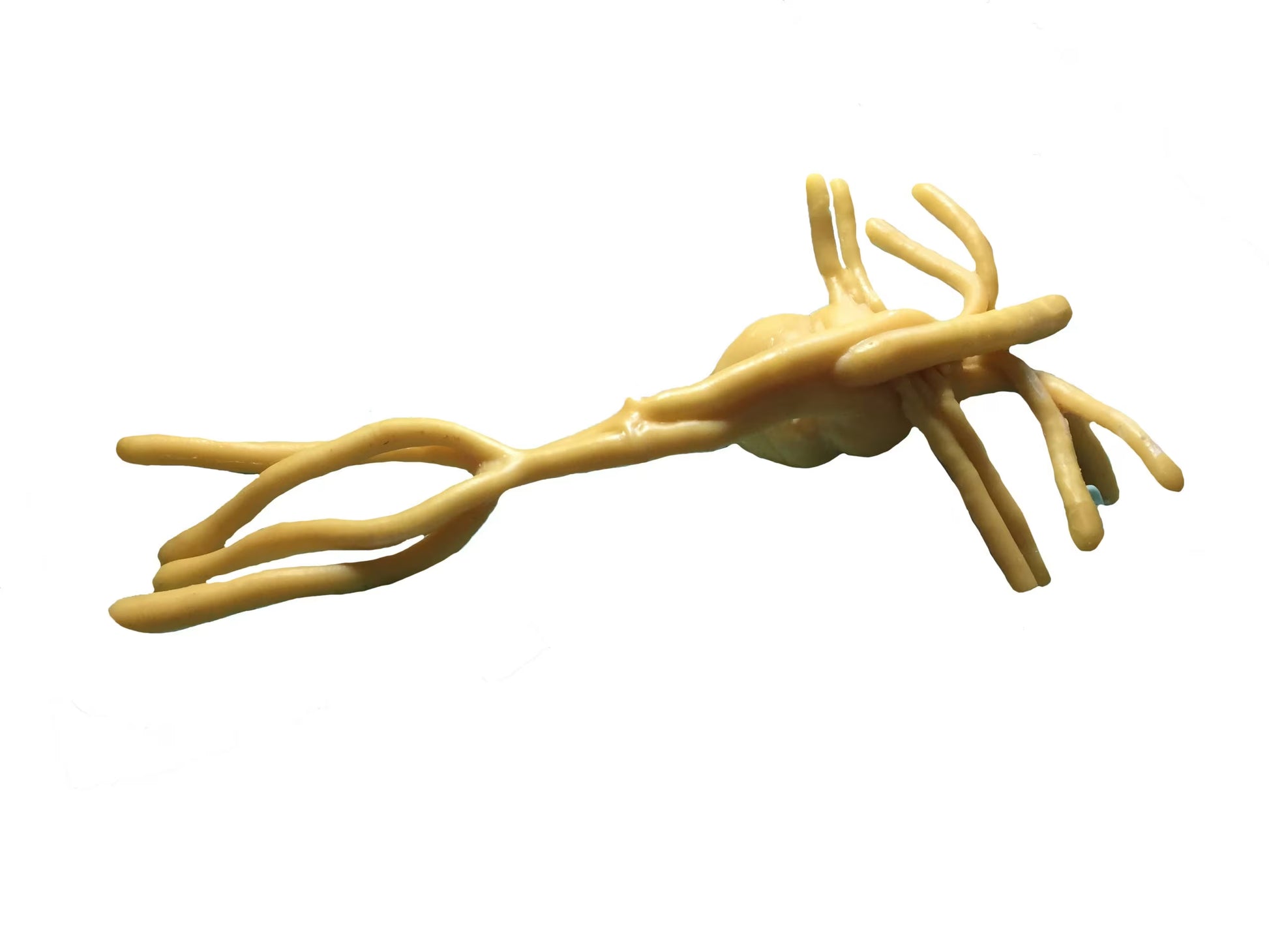
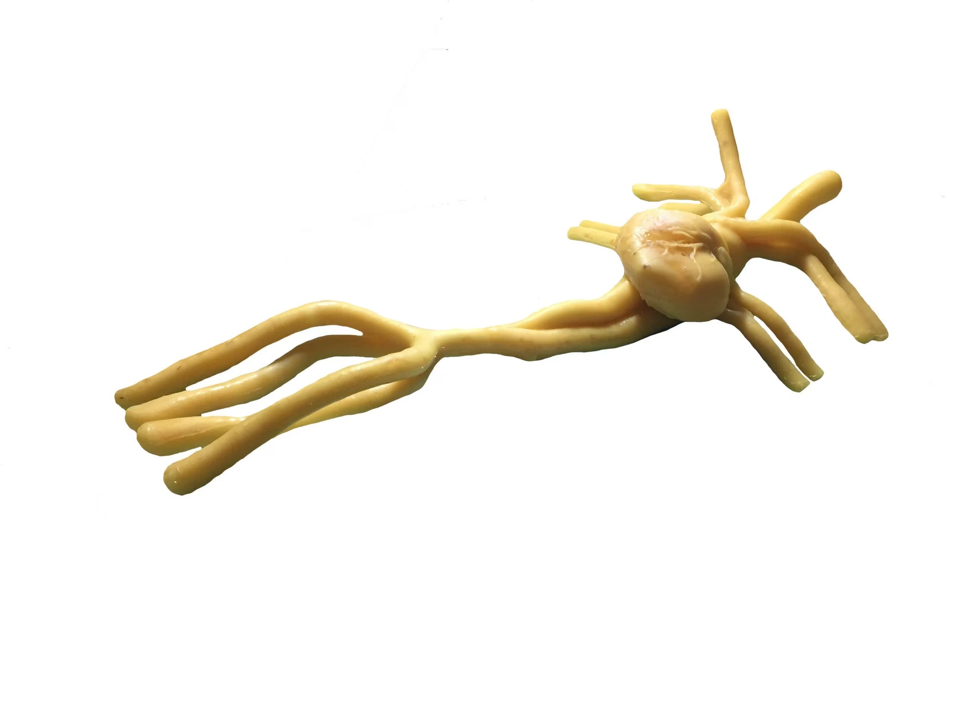
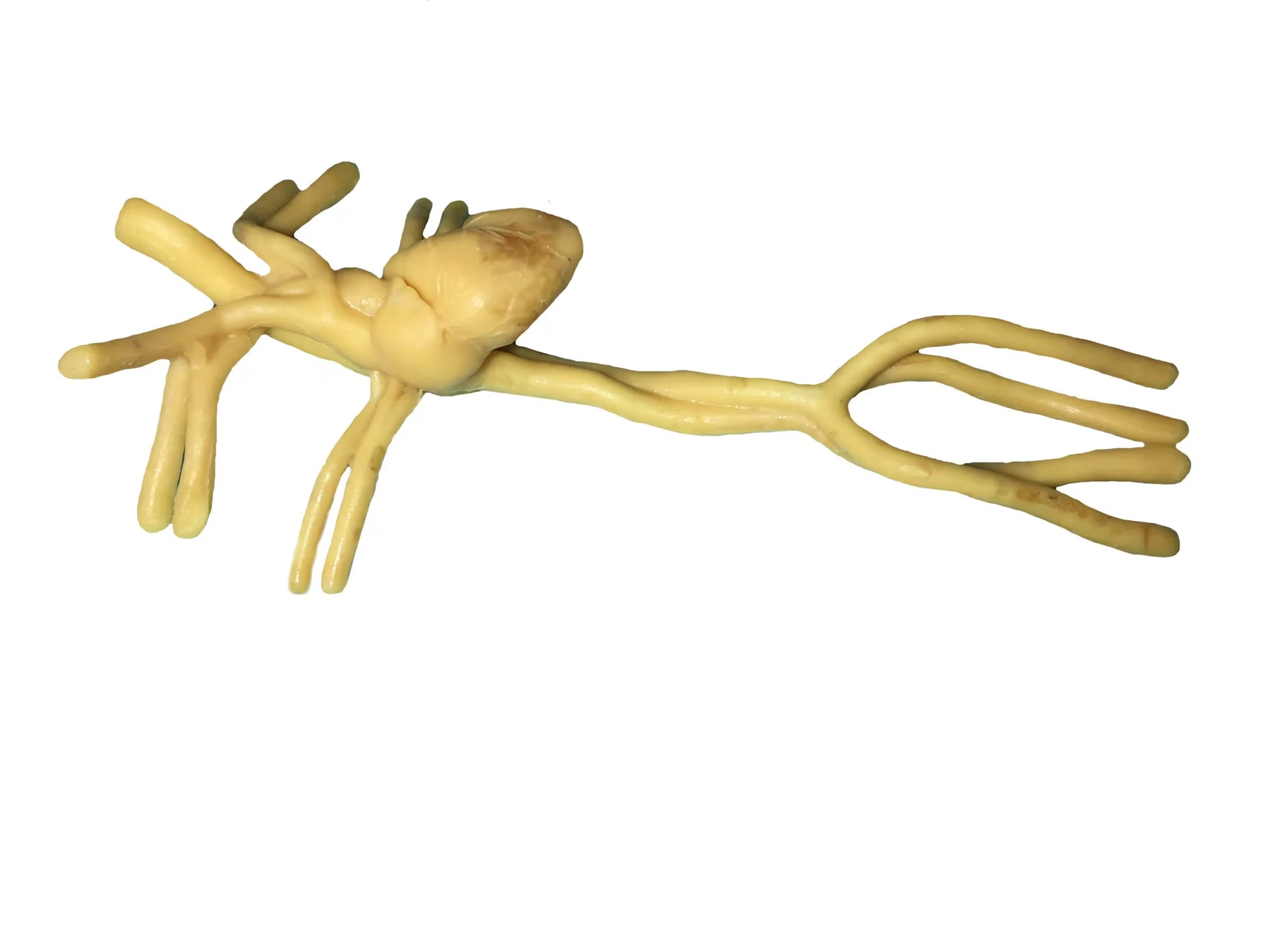
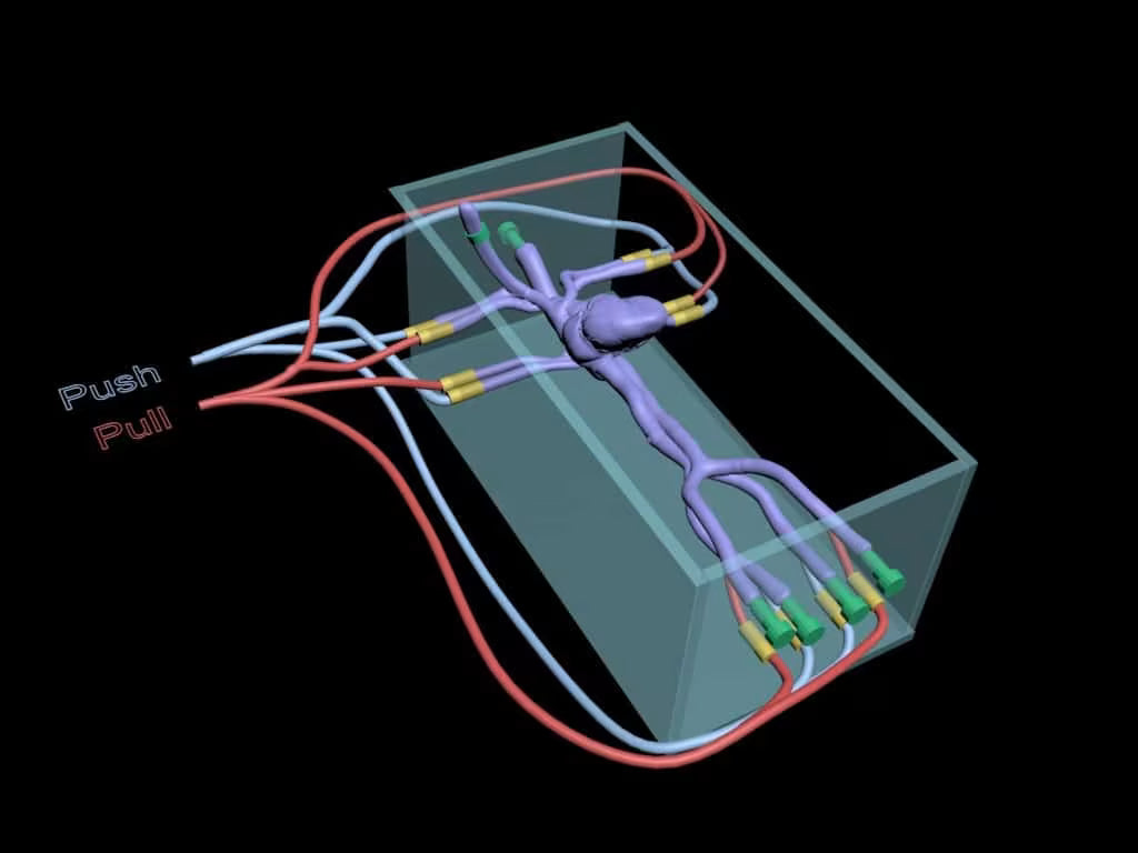
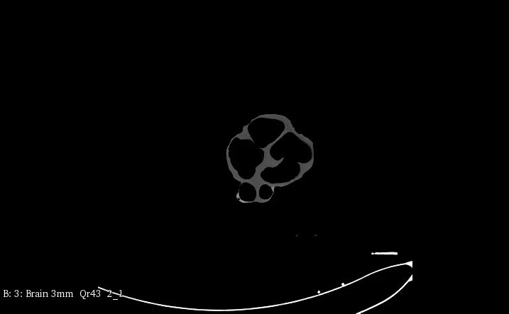
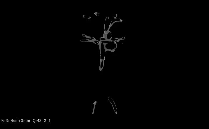
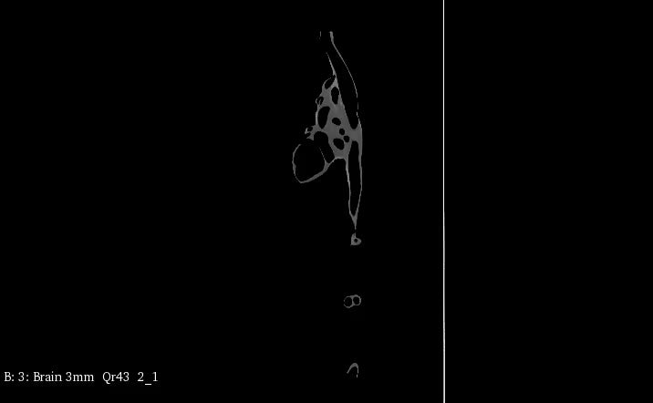
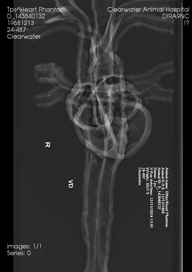

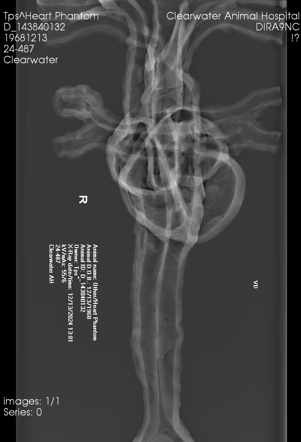
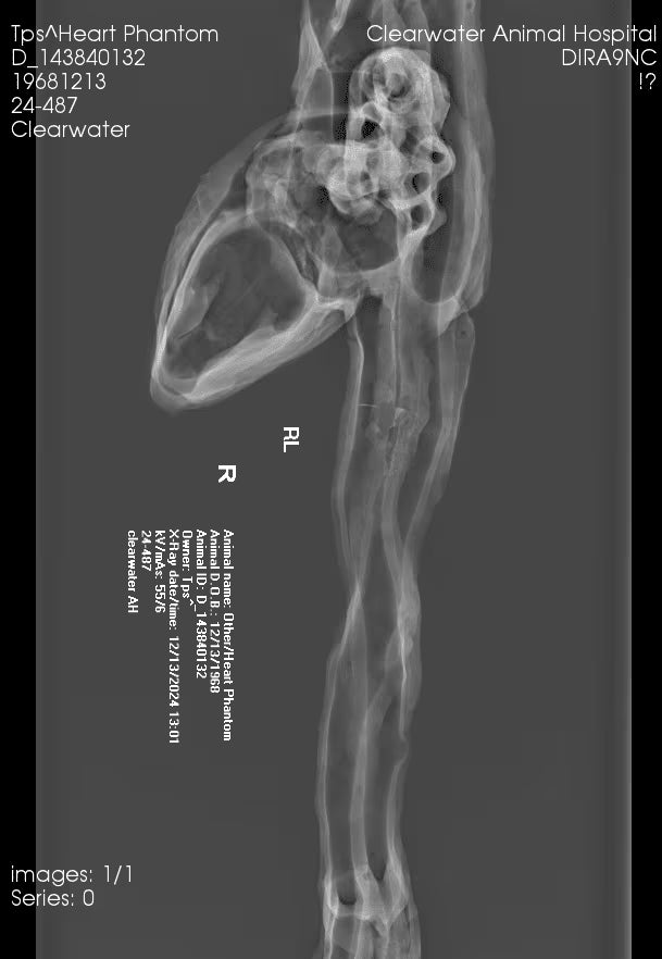
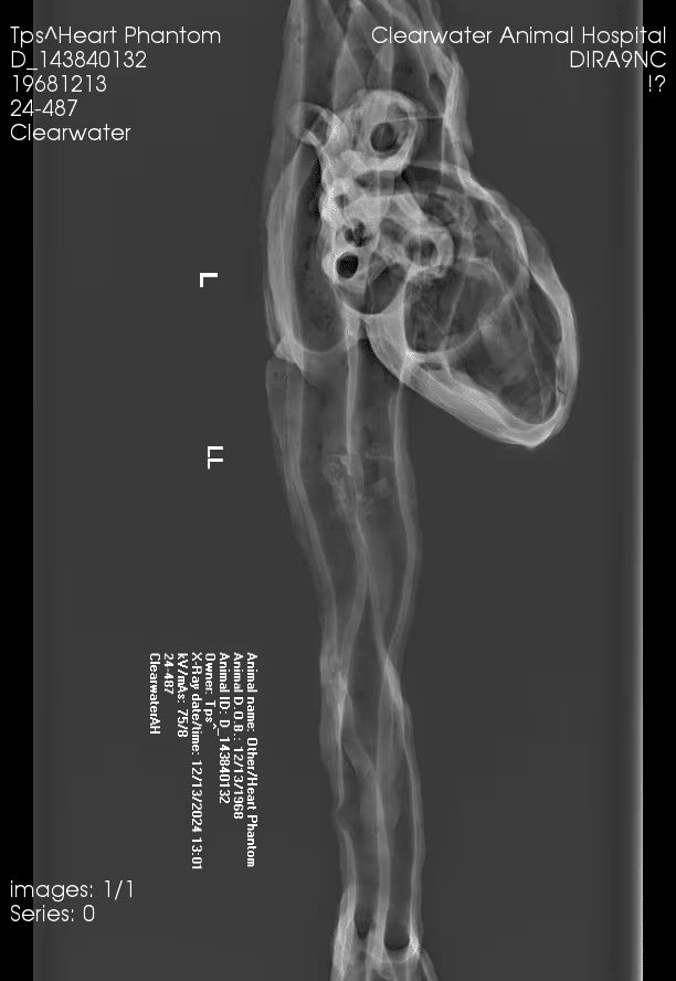
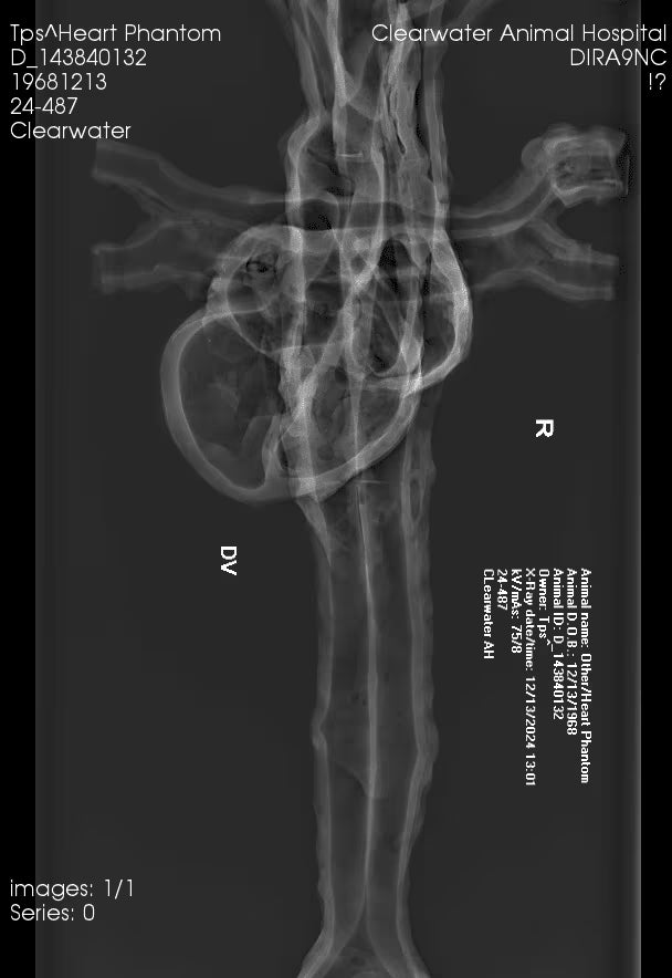
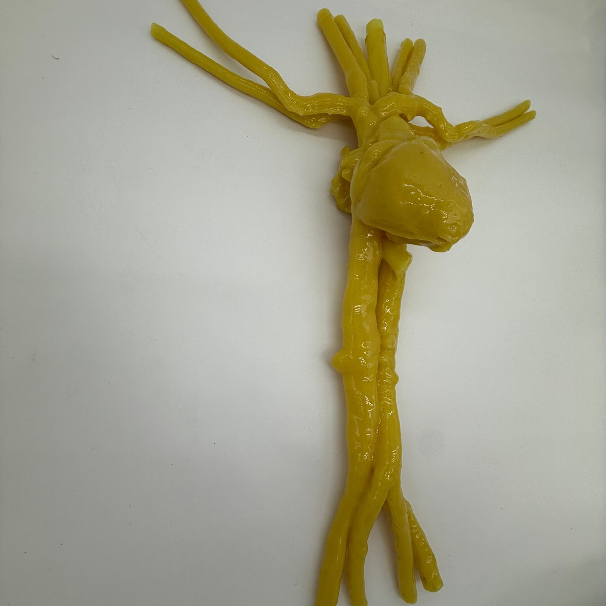
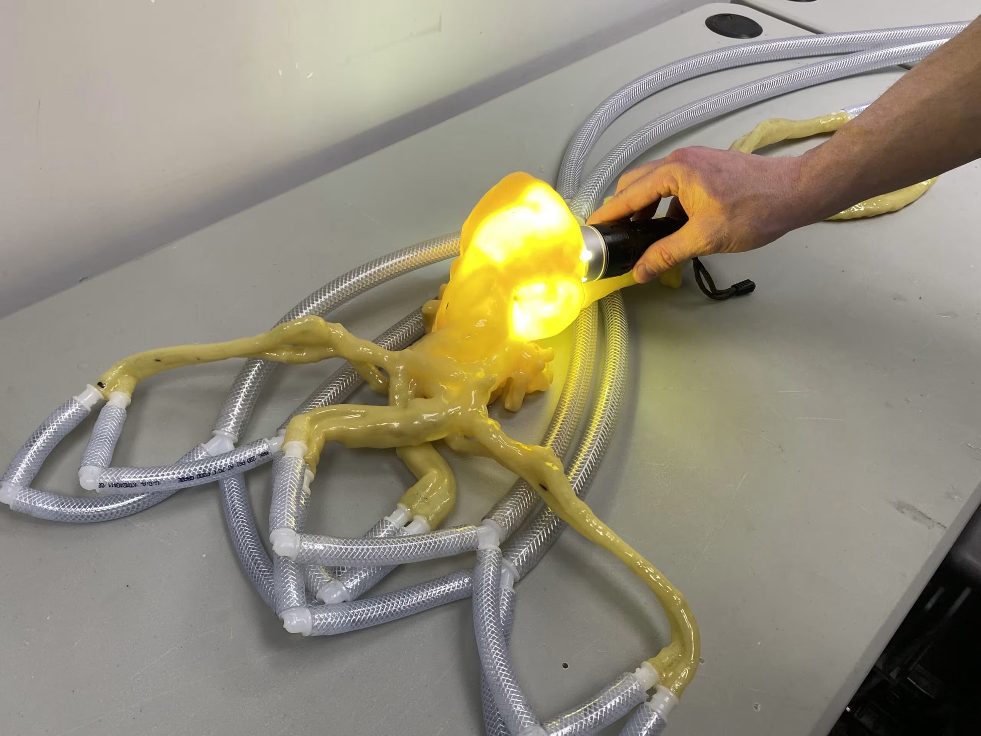
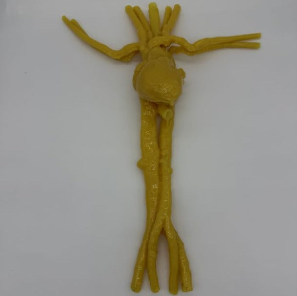
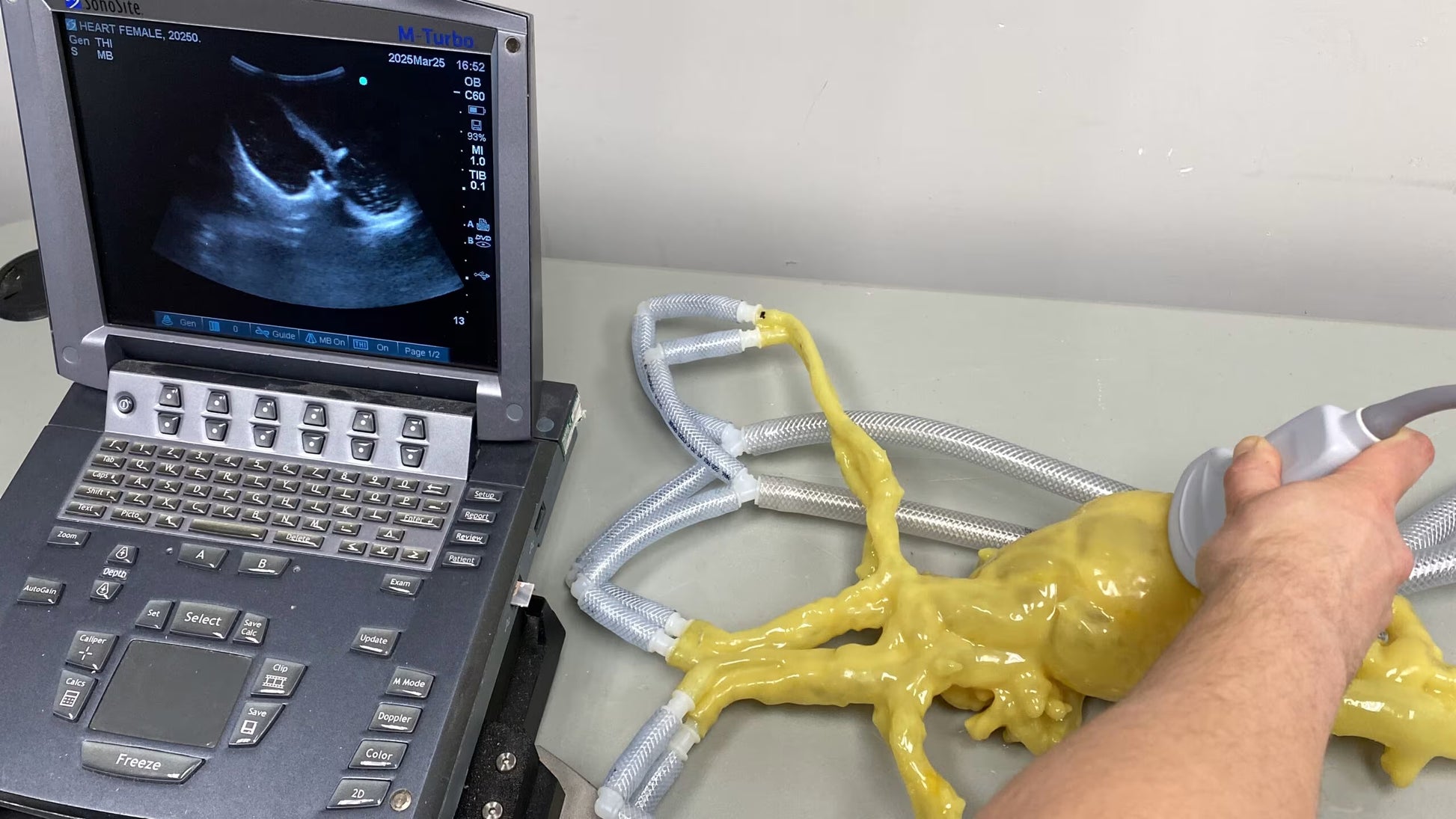
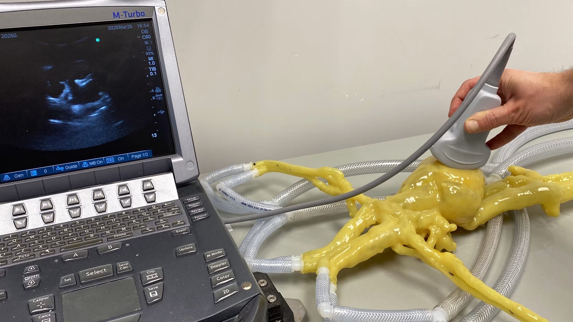
The Adult Heart Phantoms are realistic simulators of a healthy human heart, compatible with X-Ray/CT, Ultrasound, and MRI. Both male and female models support lifelike cardiac motion and can be connected to a heart pump for training and research, including echocardiograms, endoscopies, catheter insertion, angiography, and cardiovascular studies.
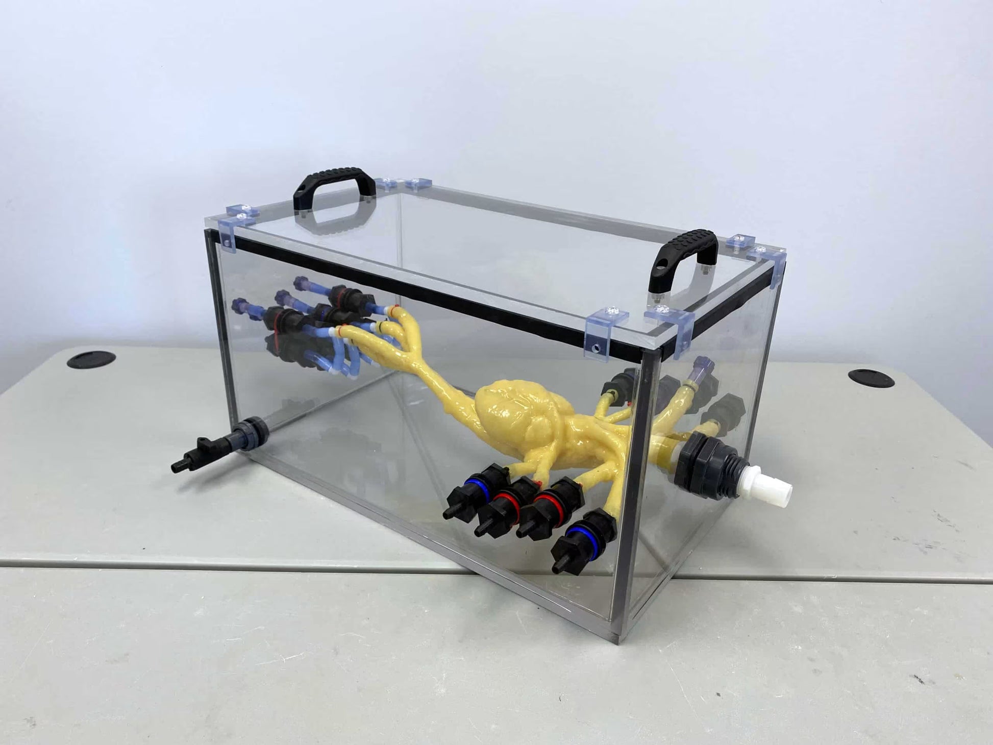
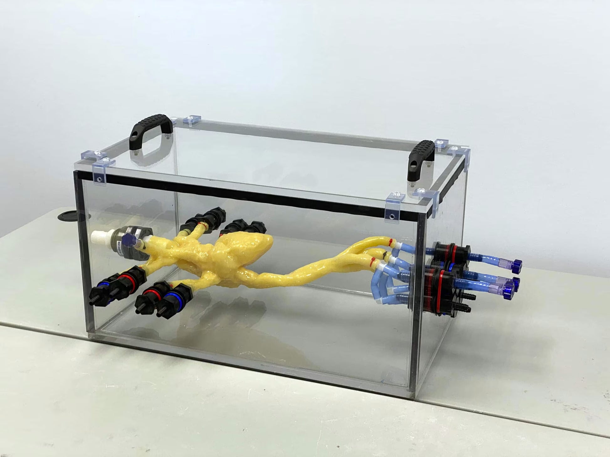
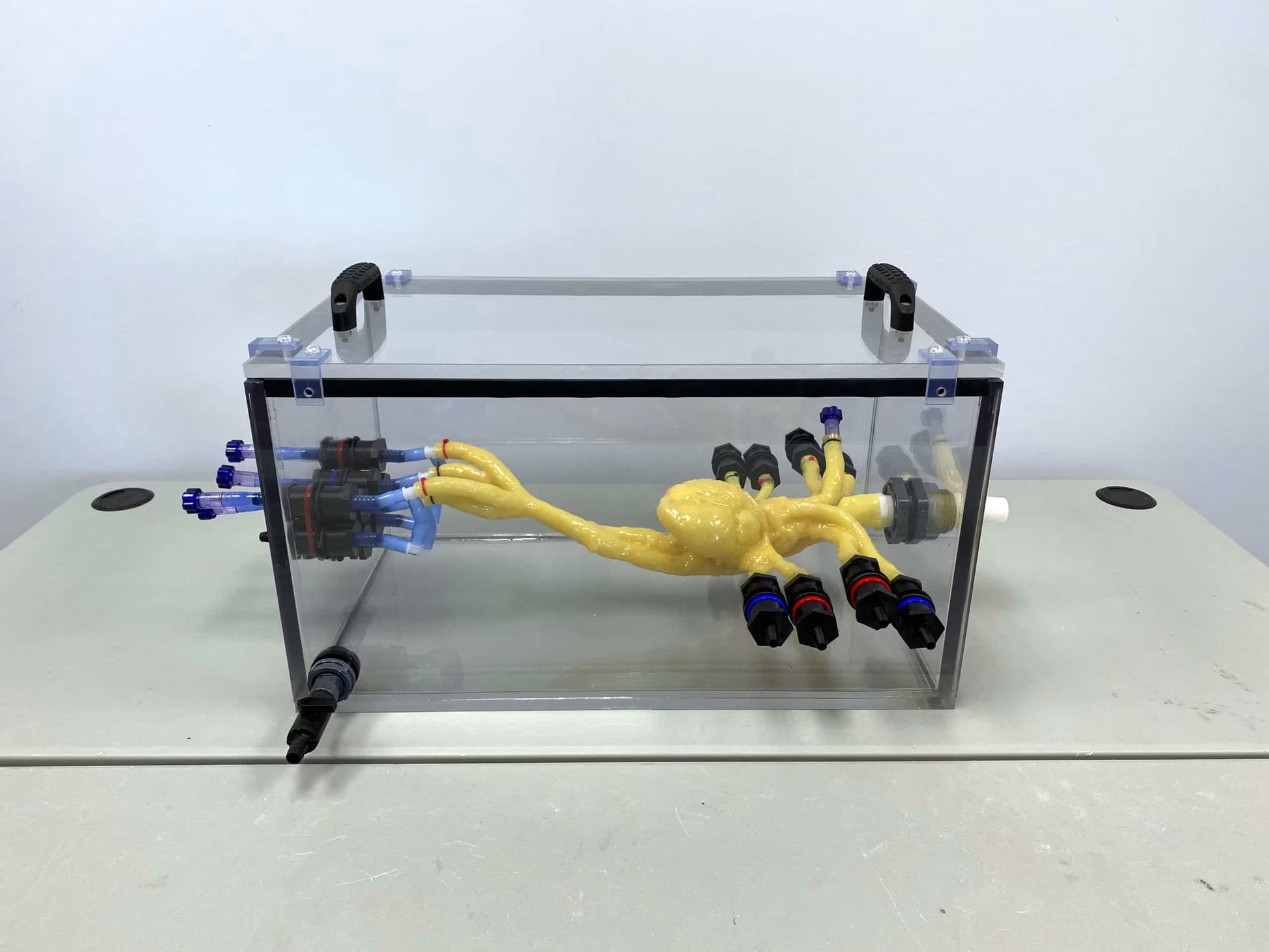
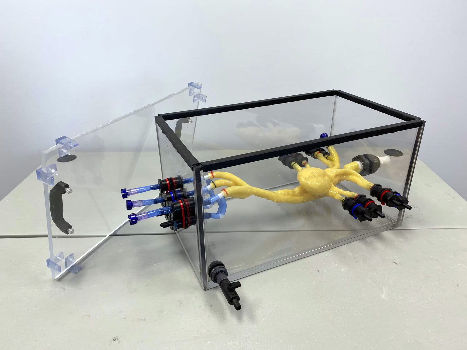
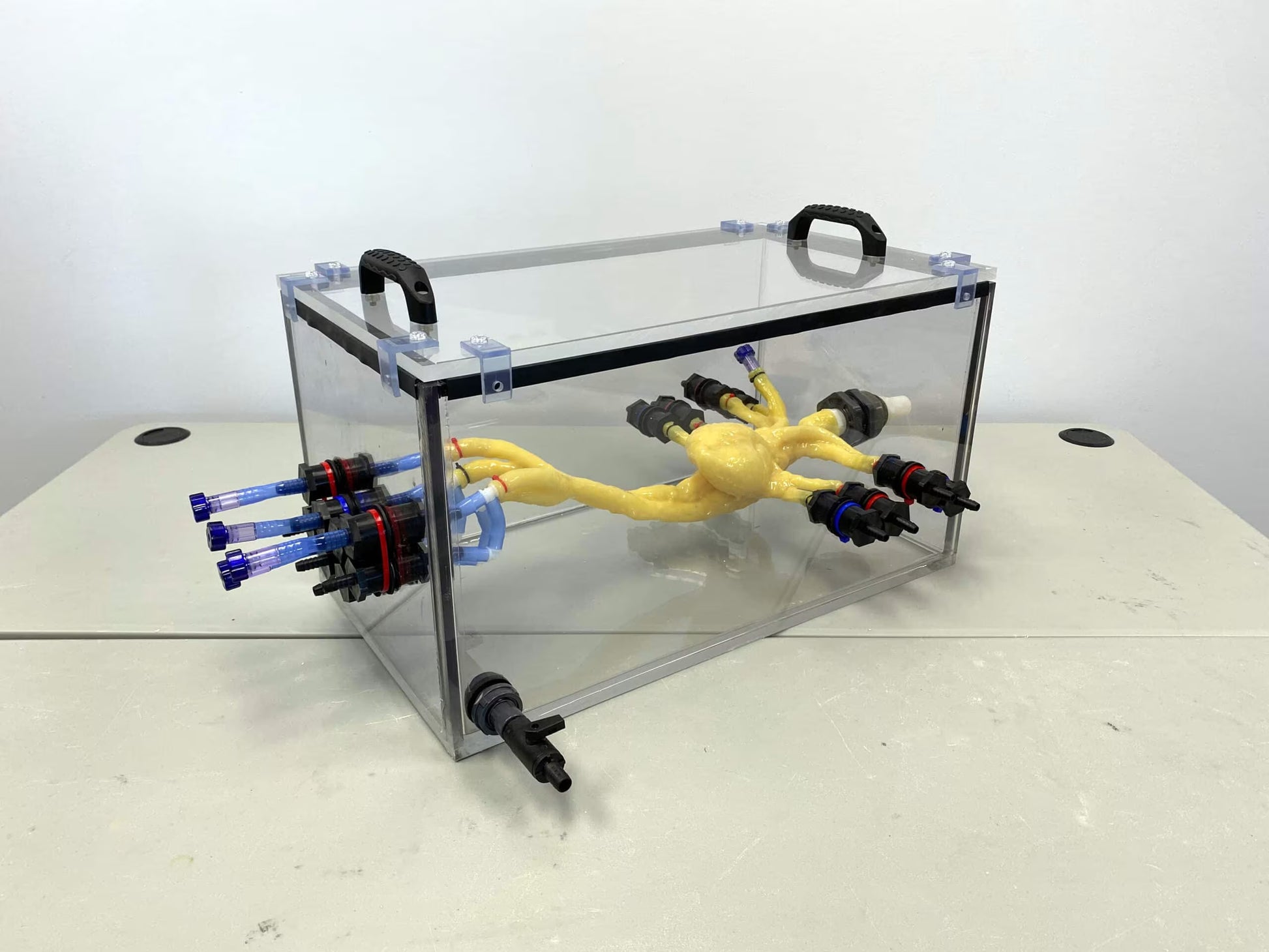
The phantoms are made from synthetic, tissue-mimicking materials that replicate the acoustical, physical, and mechanical properties of real biological tissue. Using 3D printing and advanced modelling techniques (including AI for the female model) they reproduce highly accurate anatomical structures. Each phantom includes a hollow oesophageal tube for transoesophageal echocardiography (TEE) procedures. A transparent water tank is included to ensure proper mounting and optimal visualization of internal structures and heart motion.
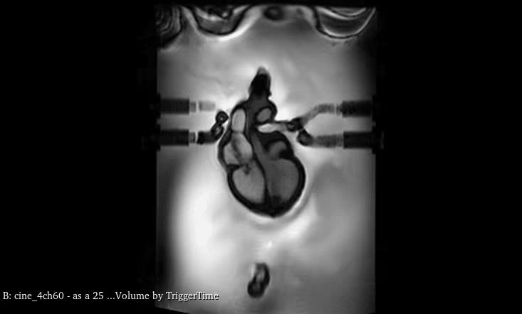
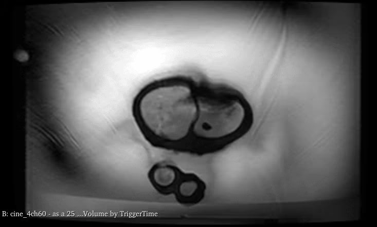
In terms of MRI applications, the tissues have realistic T2 relaxation times, making them ideal for T2-weighted imaging. Proton-density imaging also yields very good results. T1-weighted imaging is possible, though T1 values are less realistic (~100 ms).
Anatomical Features & Volumes
- Right Ventricle: Male 60 ml, Female 48 ml (Moderator Band present)
- Left Ventricle: Male 63 ml, Female 74 ml (Papillary muscles to anterior/posterior walls)
- Right Atrium: Male 45 ml, Female 52 ml (Distinct muscle structures in appendage)
- Left Atrium: Male 29 ml, Female 64 ml (Unique appendage muscle structures)
- 4 Valves
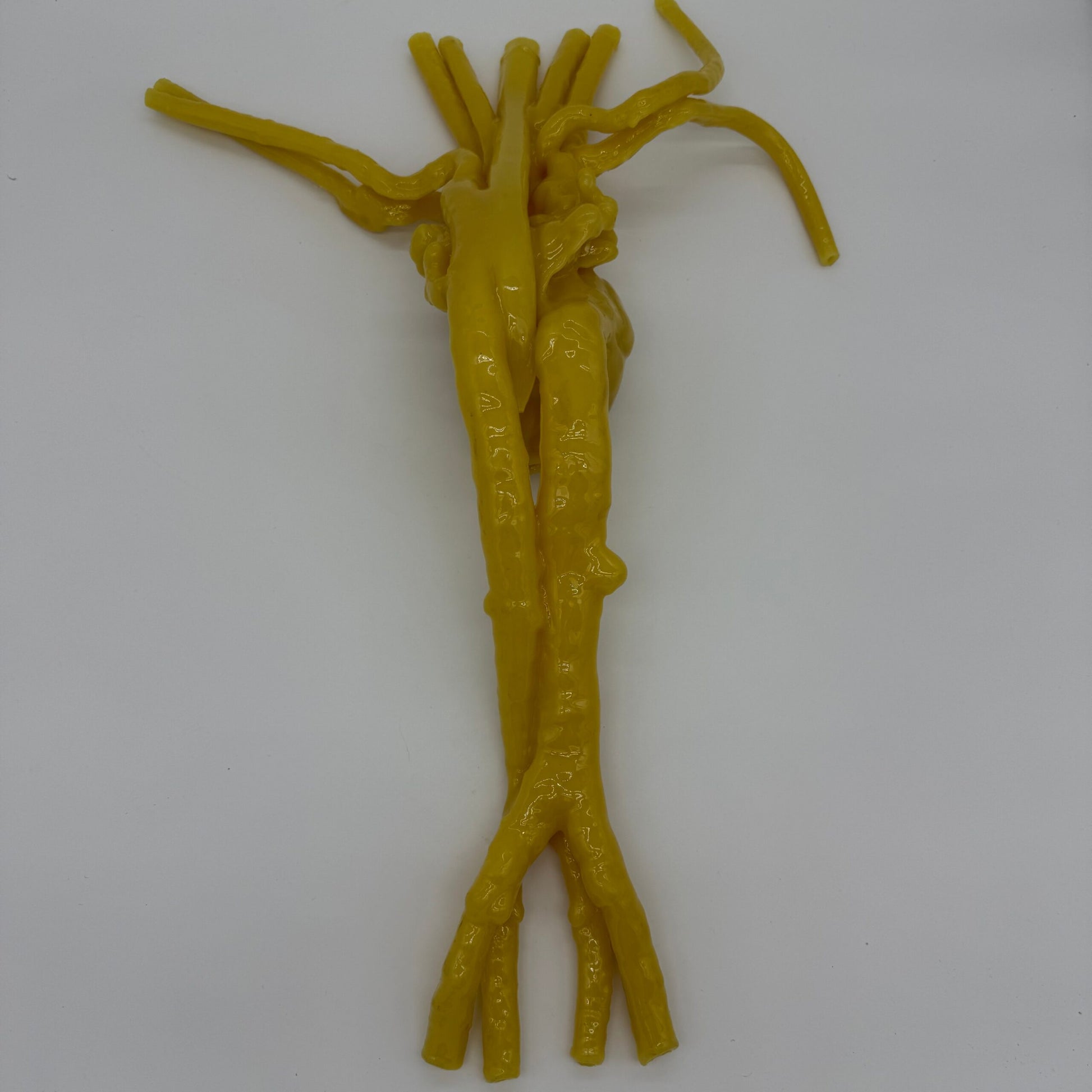
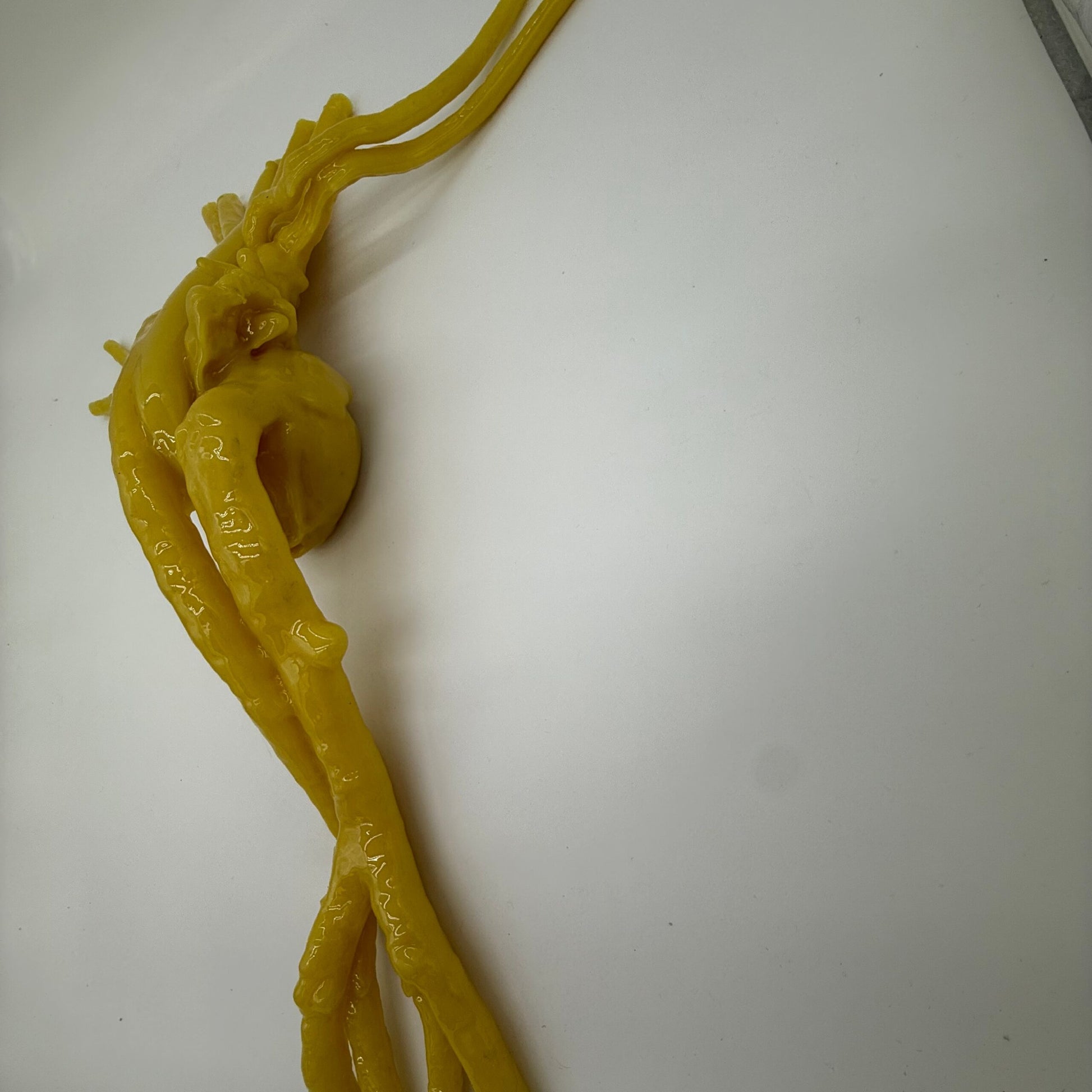
- SVC, IVC, Aorta
- Arteries: Right & Left Subclavian, Right & Left Pulmonary (Male) / Carotid (Female), Right & Left Femoral
- Veins: Right & Left Subclavian, Right & Left Pulmonary (Male) / Jugular (Female), Right & Left Femoral
- Appendage Feature
- Flexible Valvular Structures
- Oesophagus
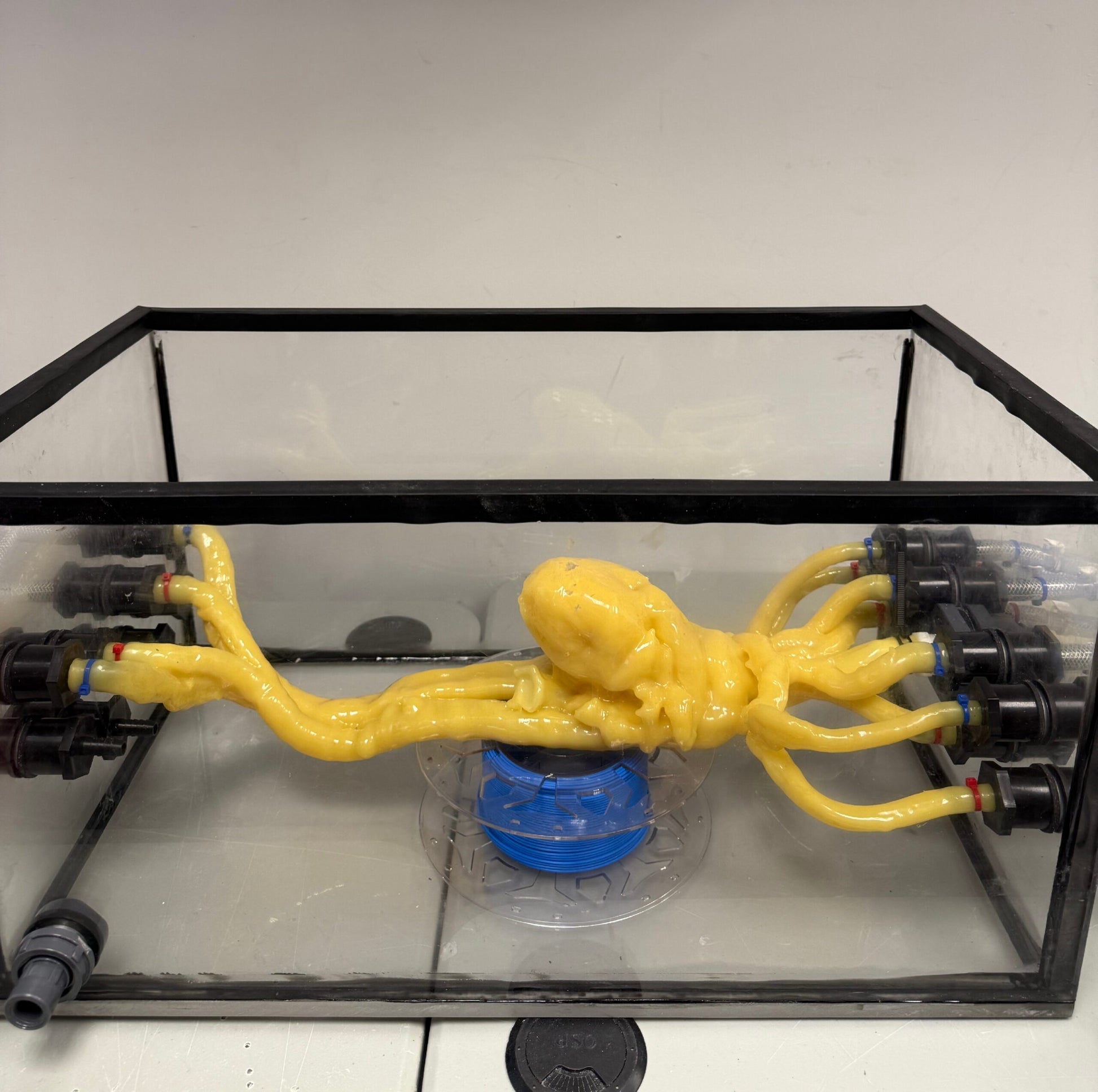
- Placed inside transparent water tank with access points
- Soft tissue and organs made using urethane-based soft resin
Included:
- Adult Male or Female Heart Phantom
- Water Tank for Heart Phantom
- User Manual / Assembly Instructions
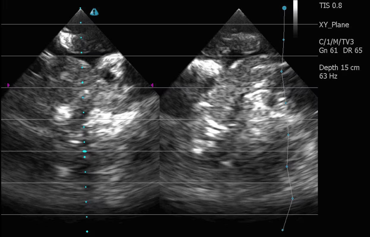
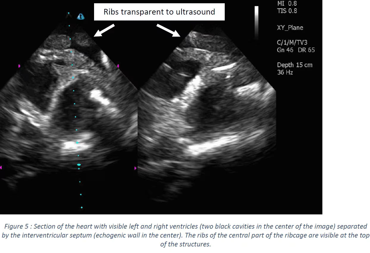
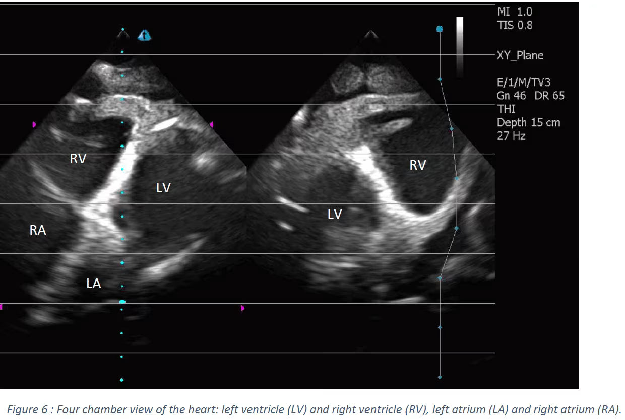
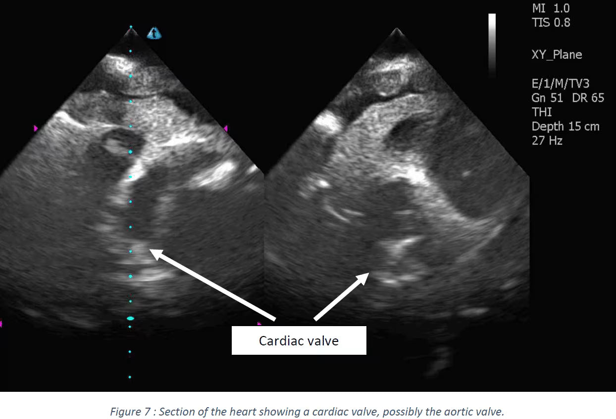
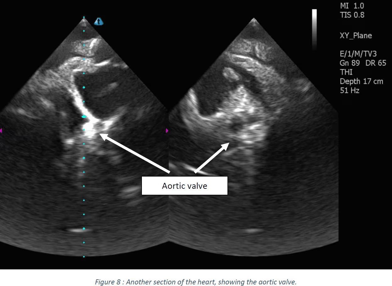
Note: Ultrasound scan images were obtained from the Adult Torso (Cardiac/FAST) [TO-A02] Phantom. The female heart anatomy was designed using AI from a CT scan of a healthy 35-year-old female patient. Male and female volumes differ slightly to reflect anatomical differences.