


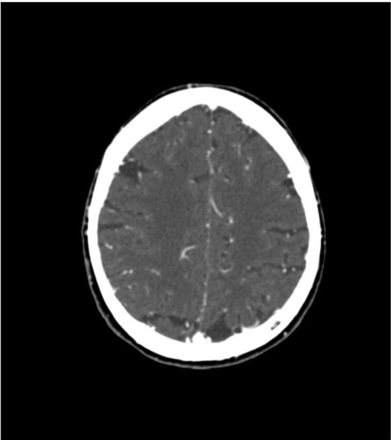
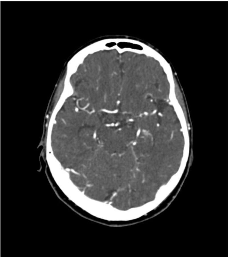
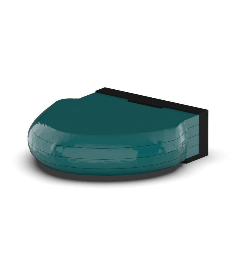
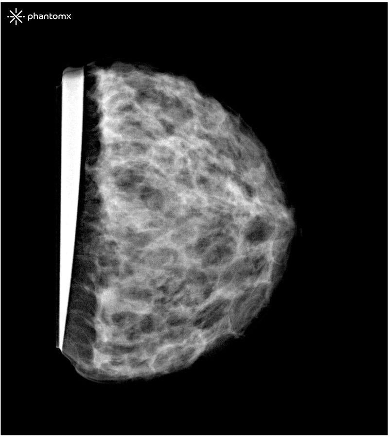
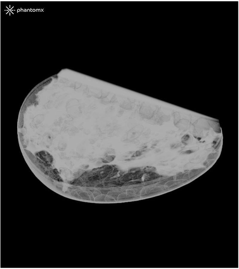
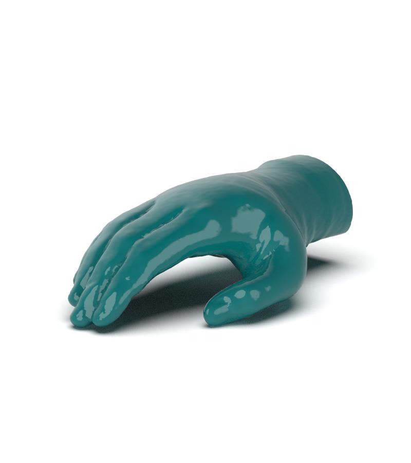
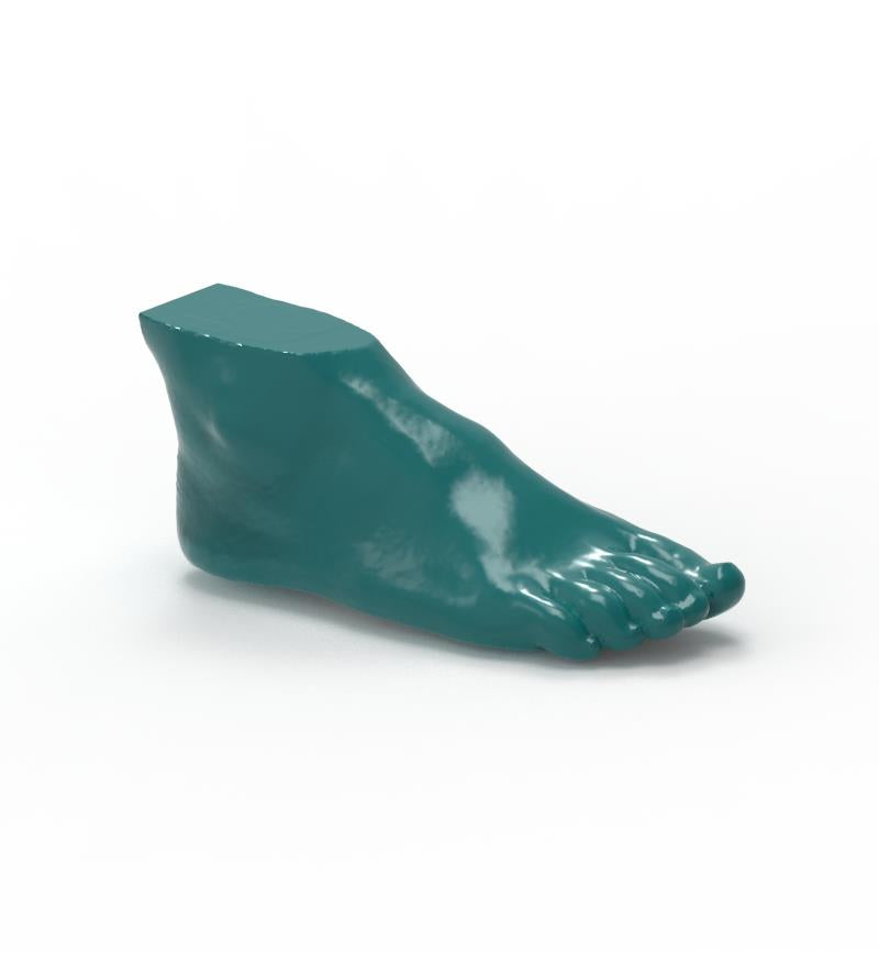
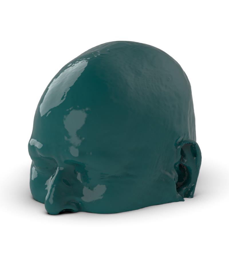
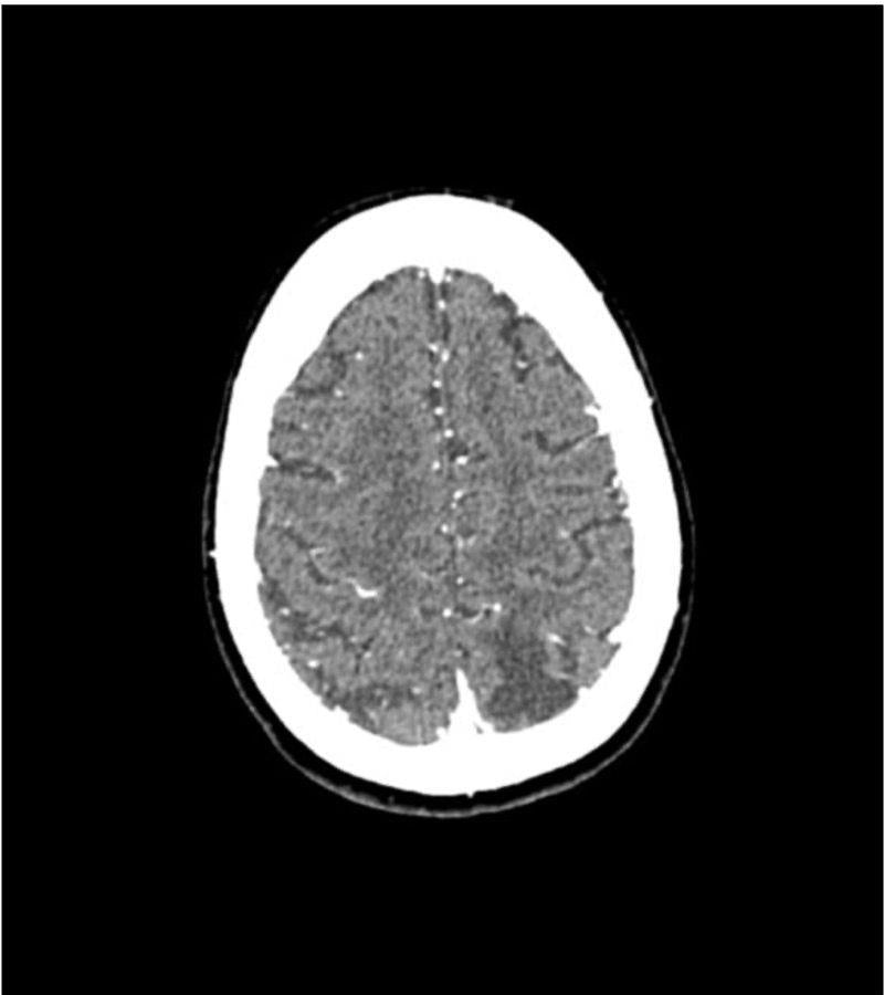
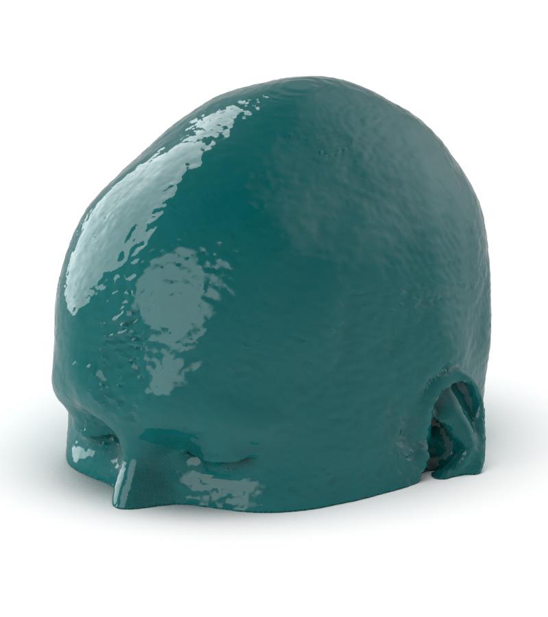
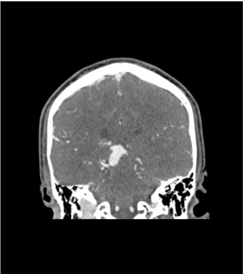

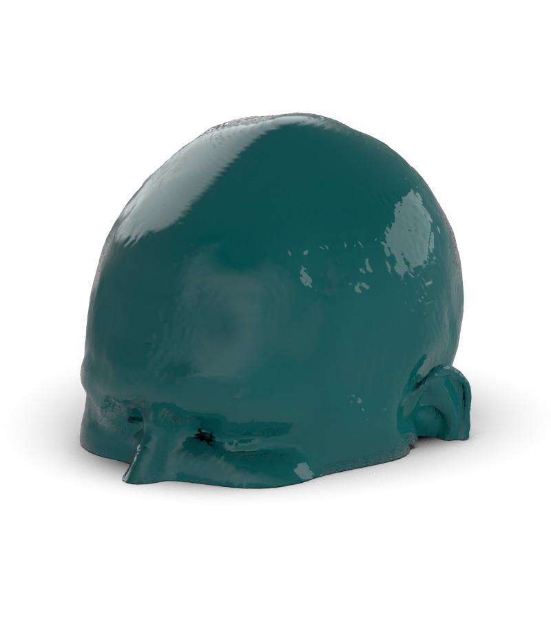
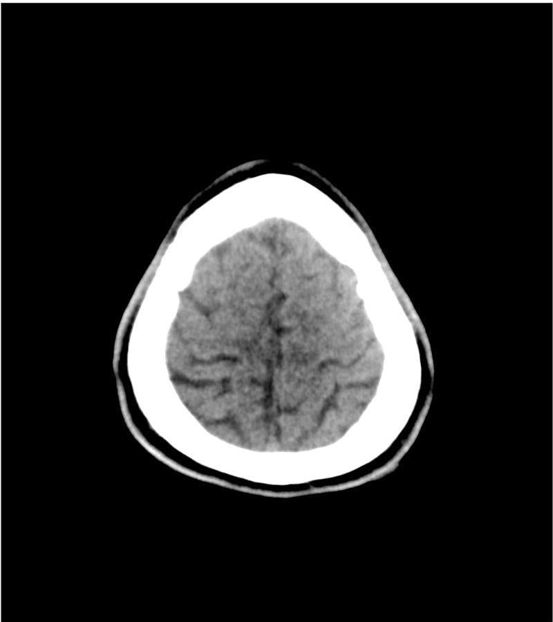
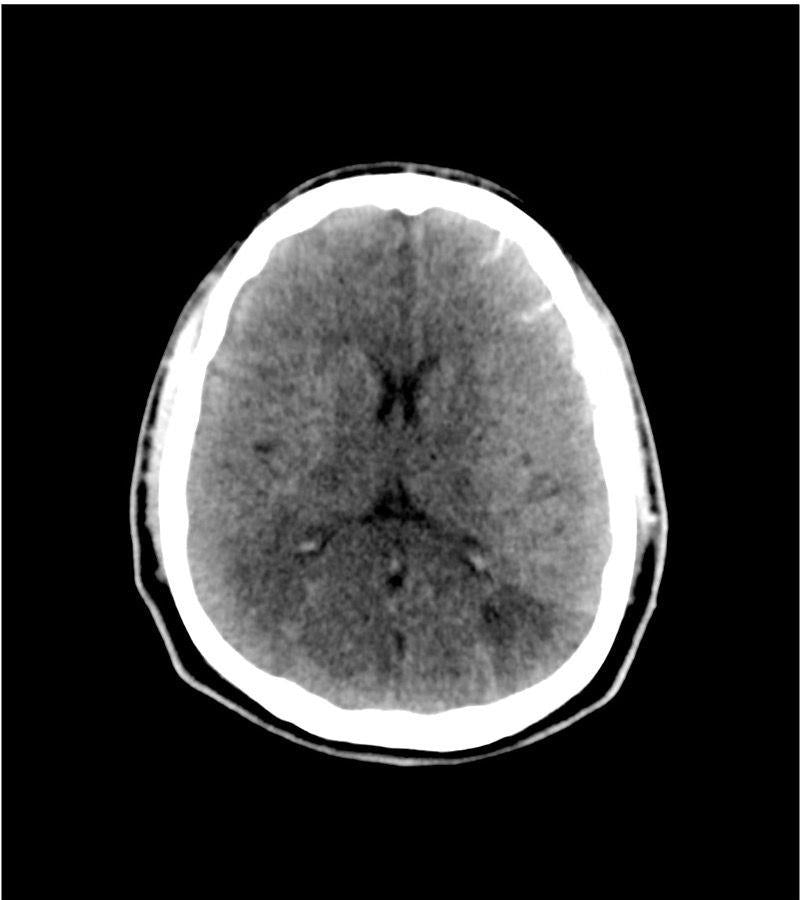
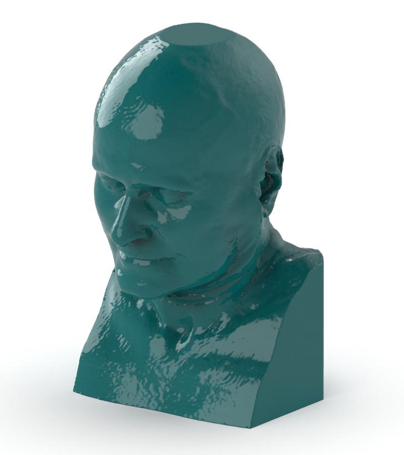
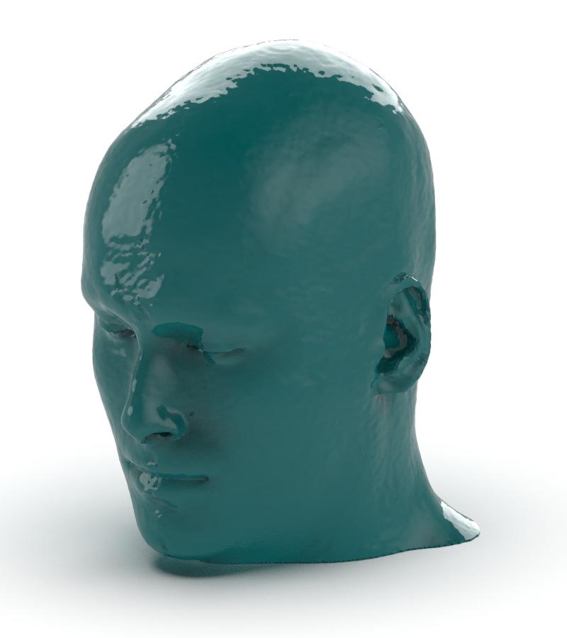
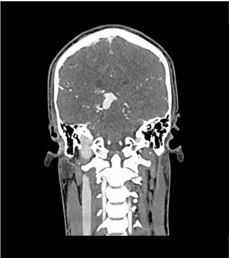
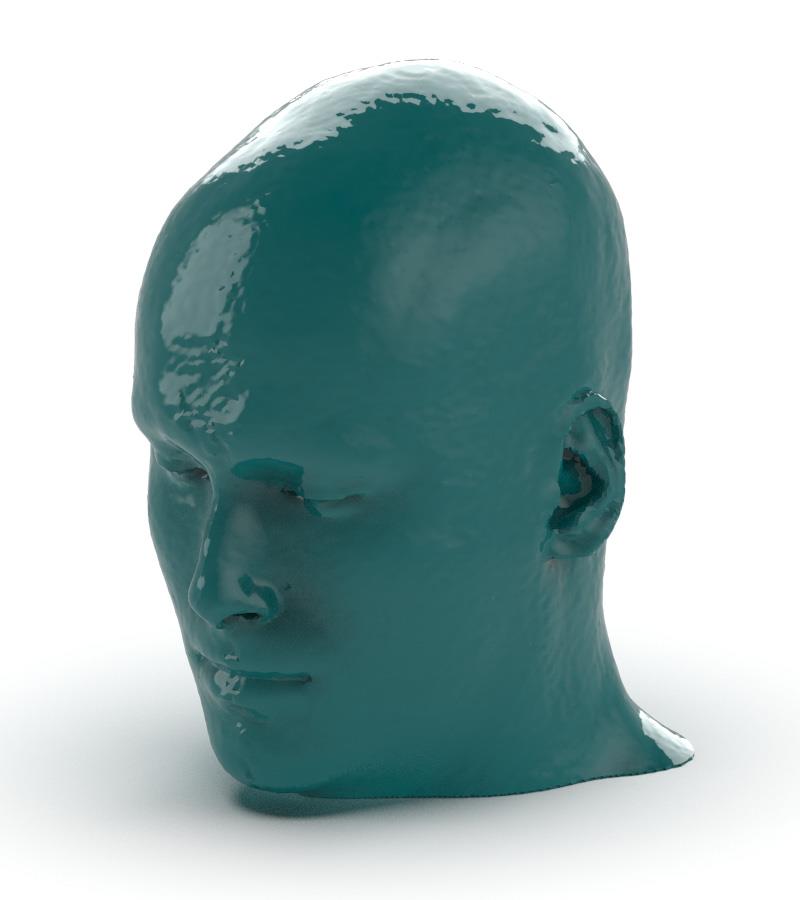

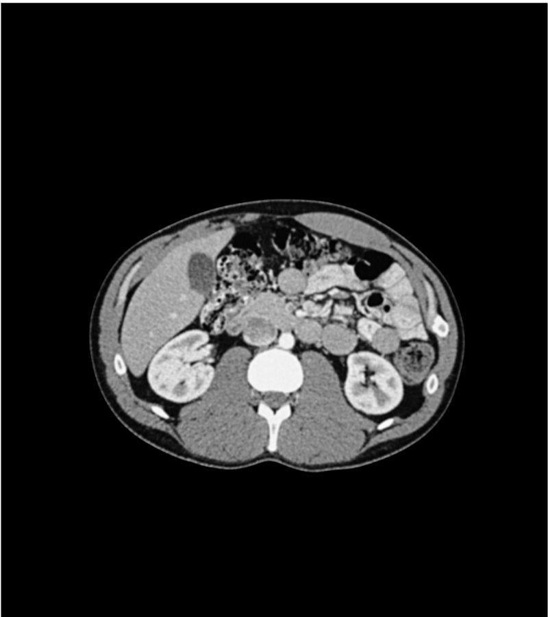

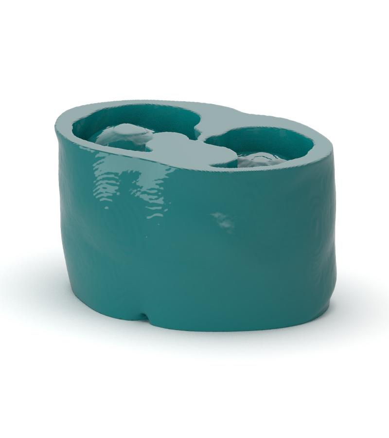
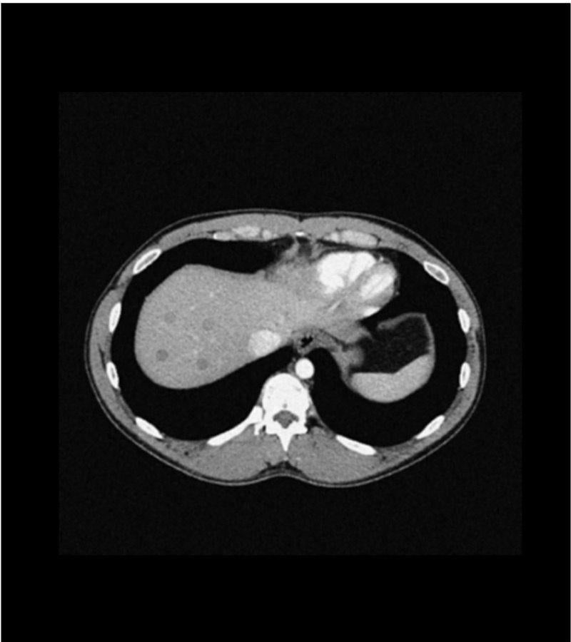
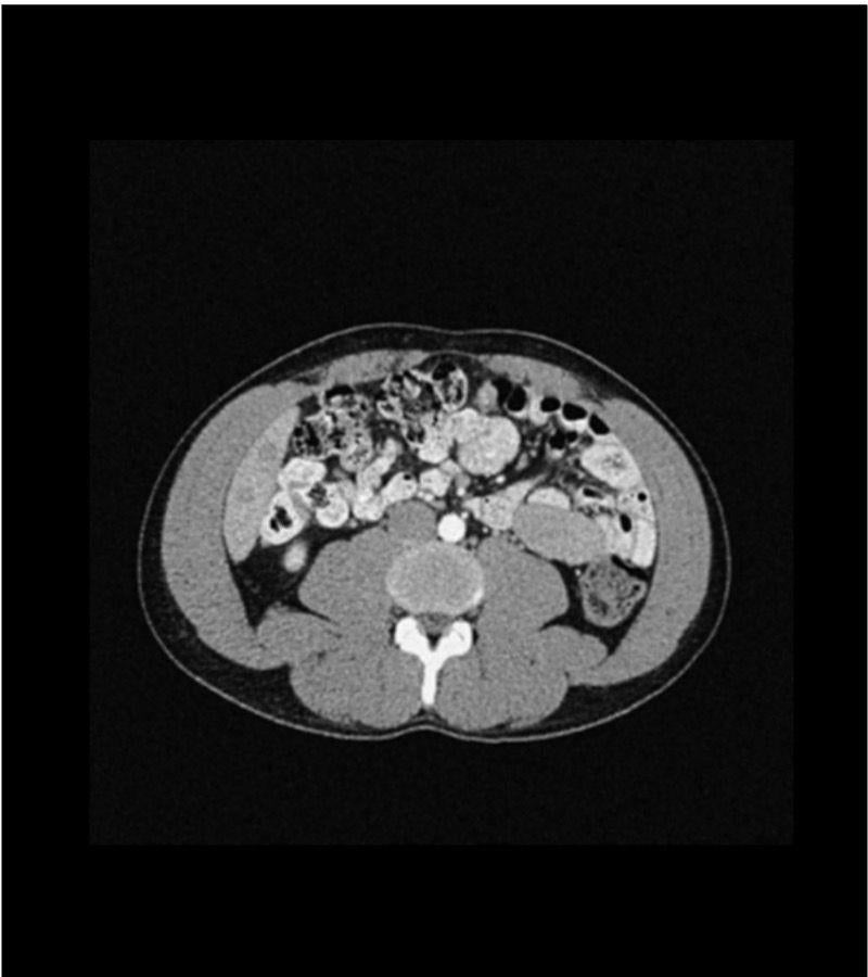
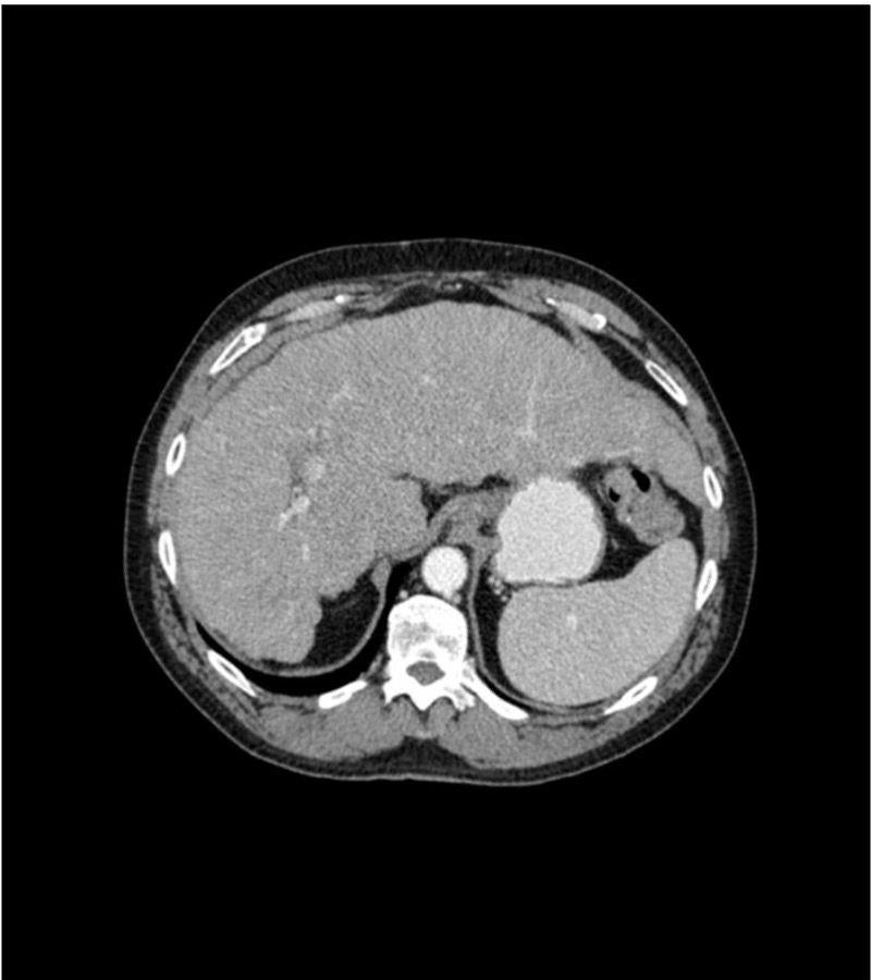
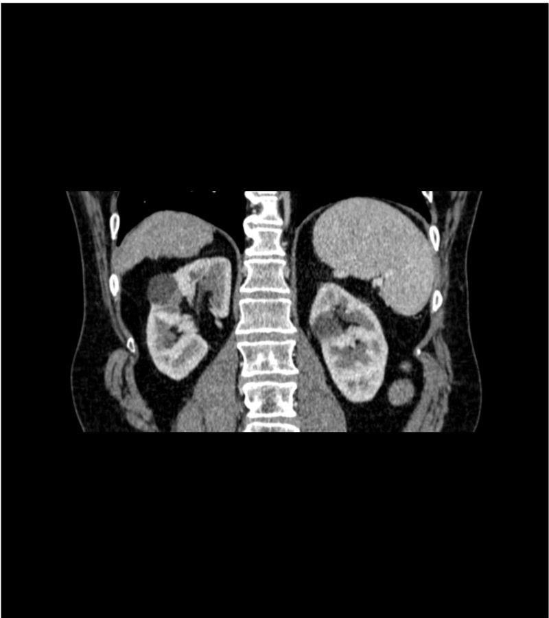
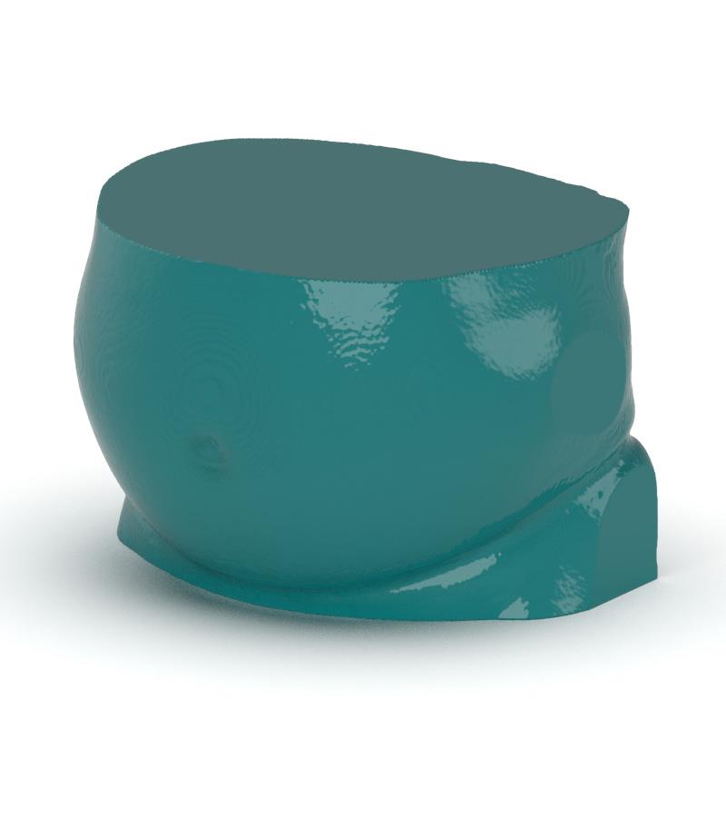
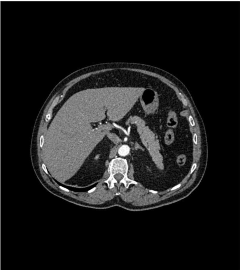
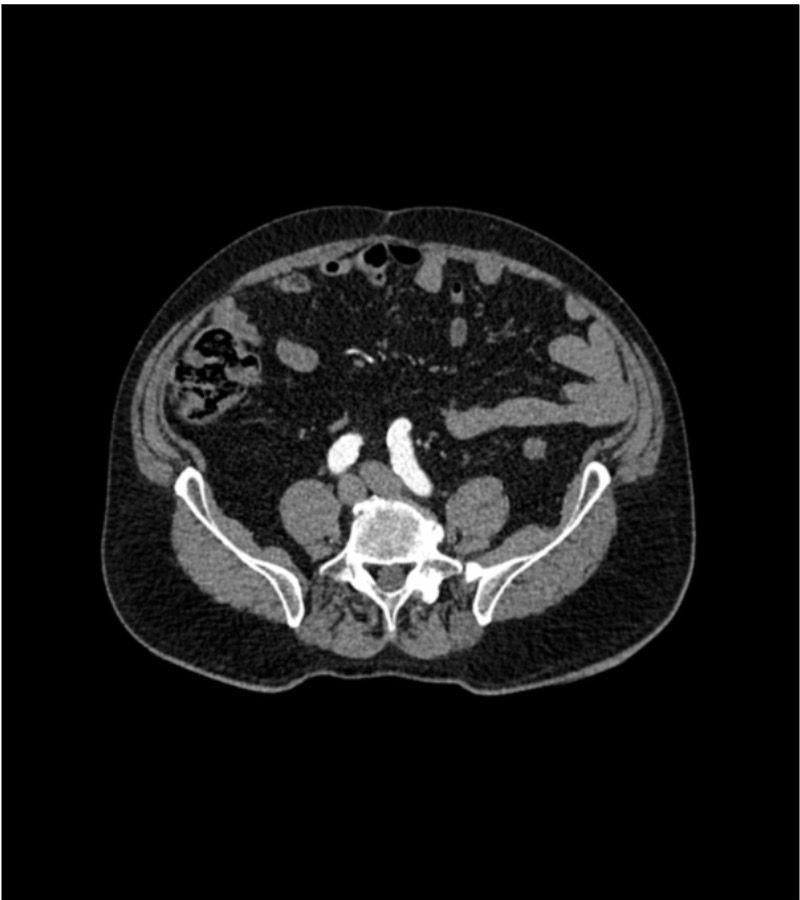

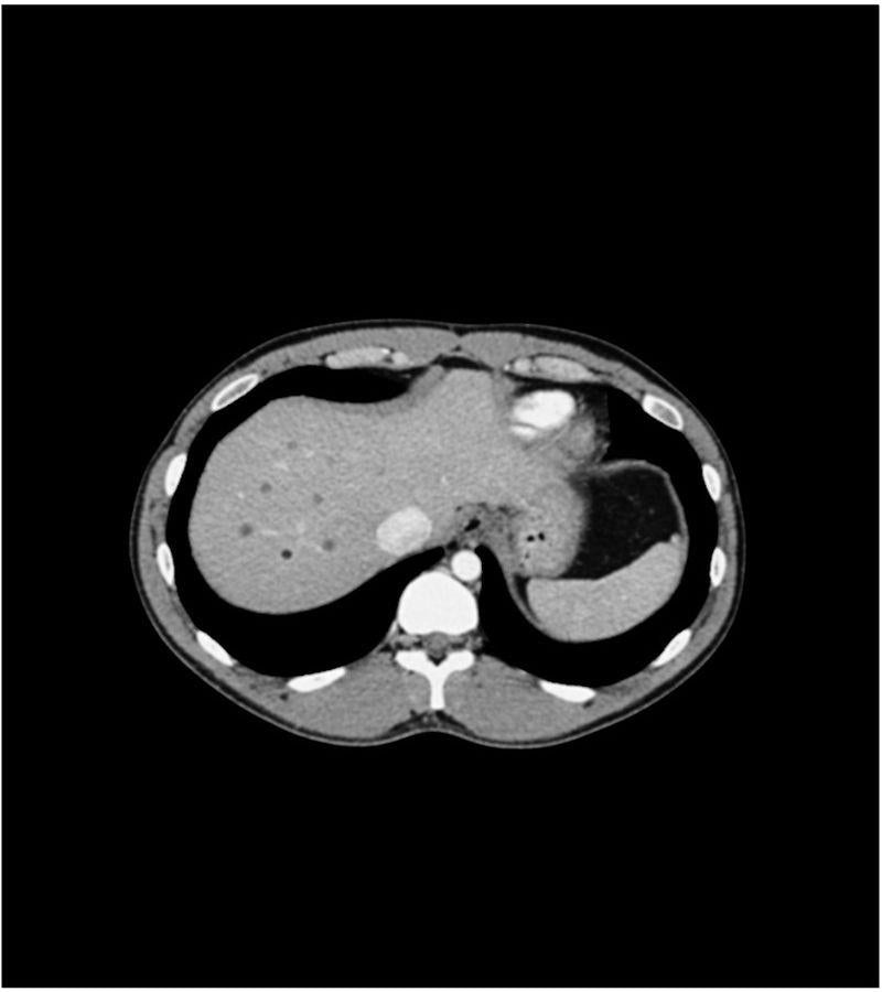
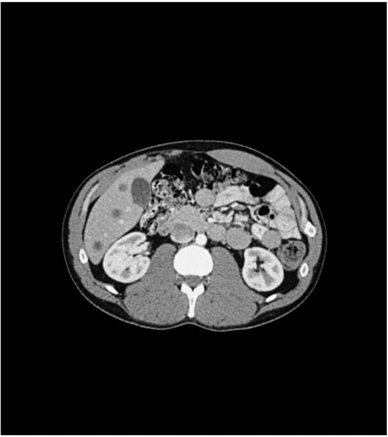
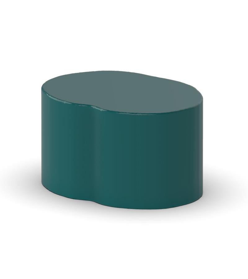
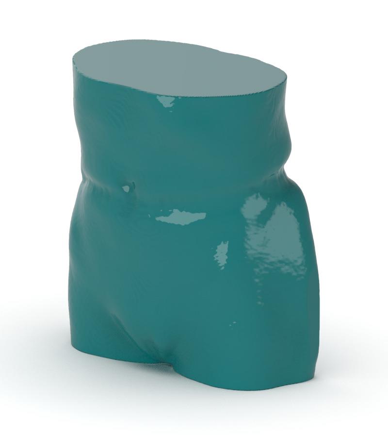
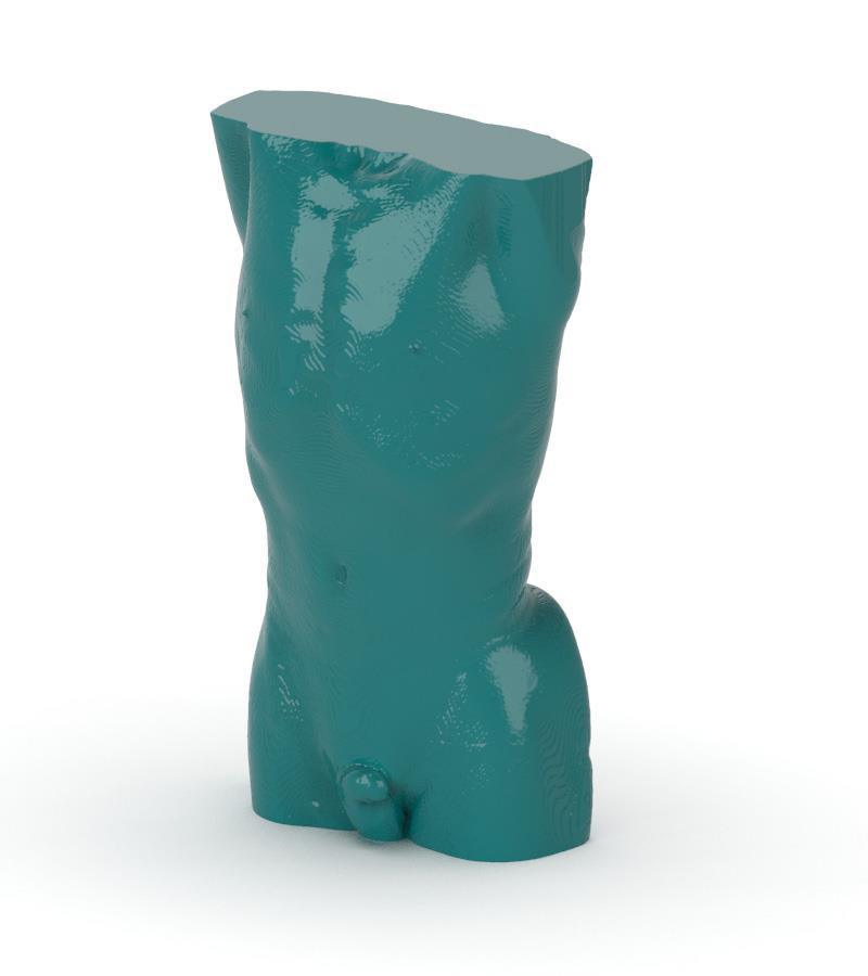
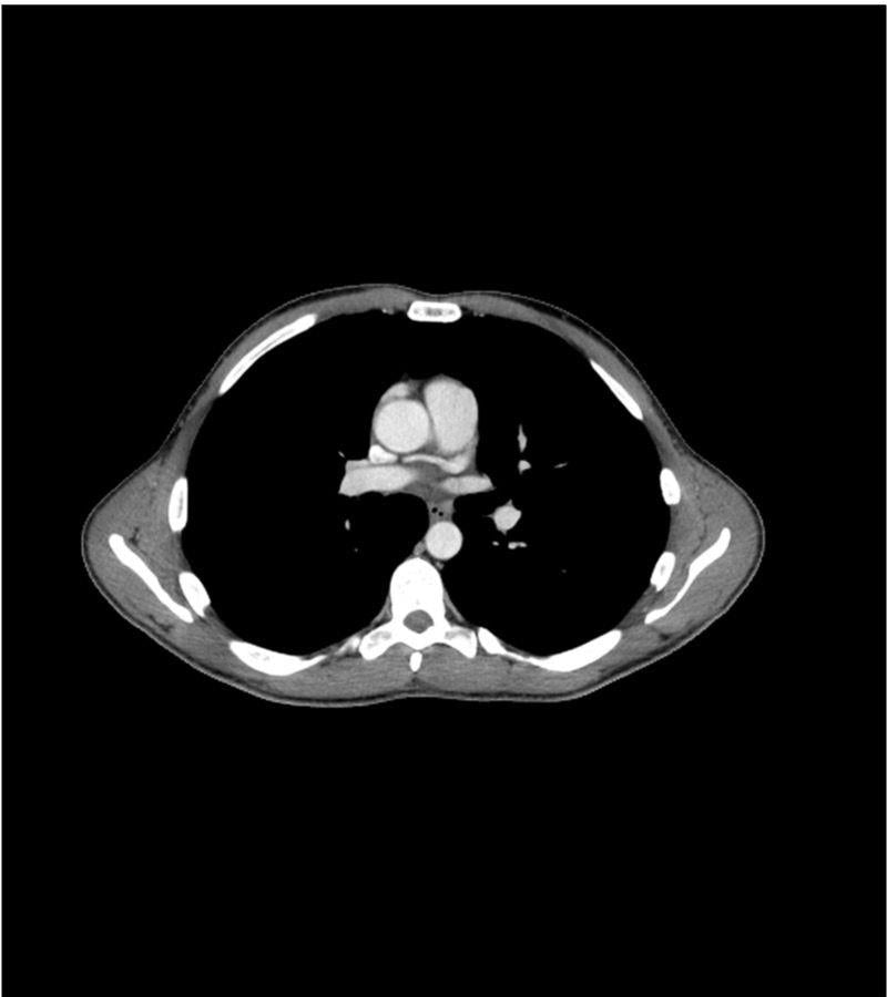
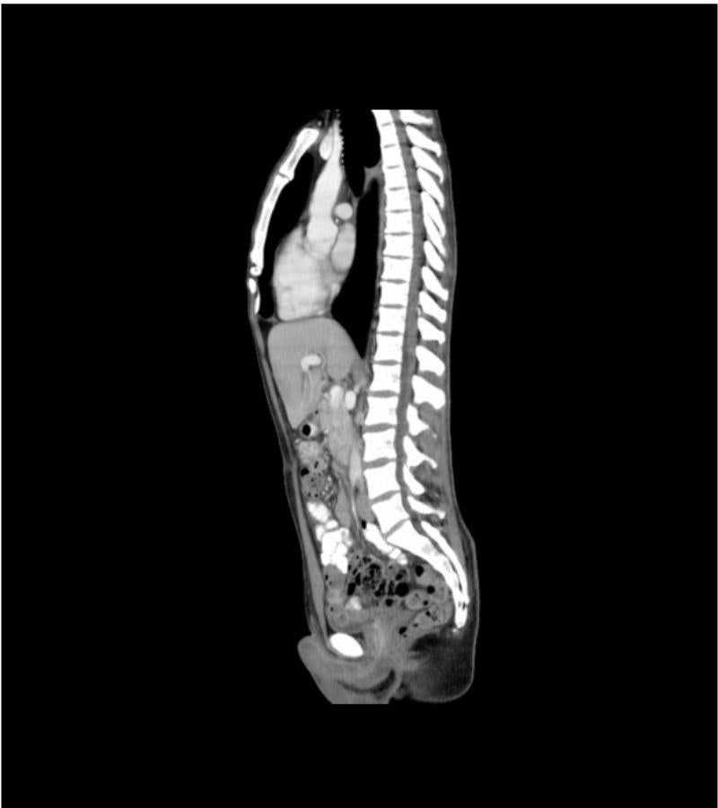
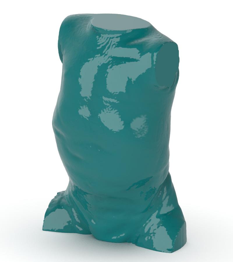
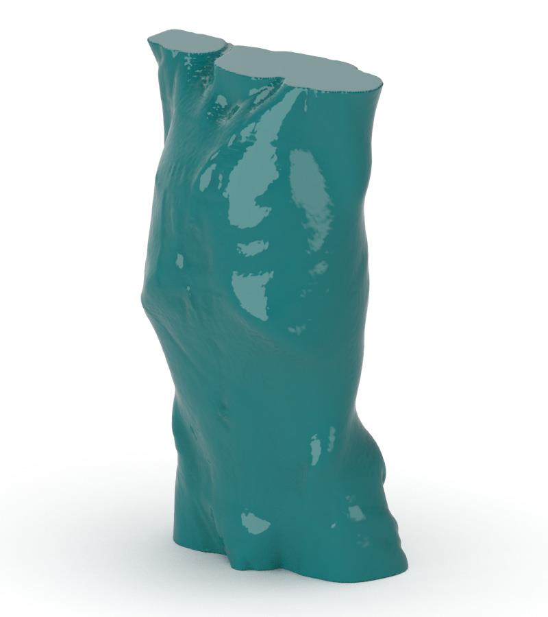
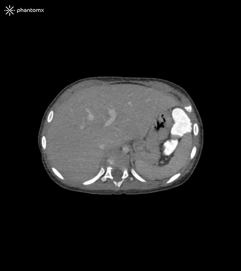
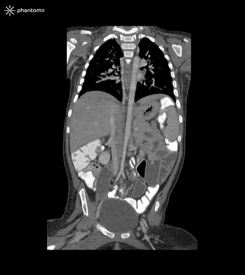
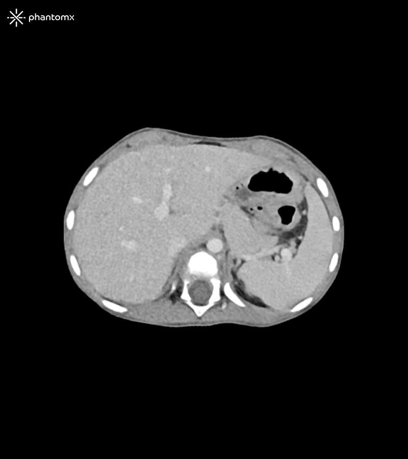
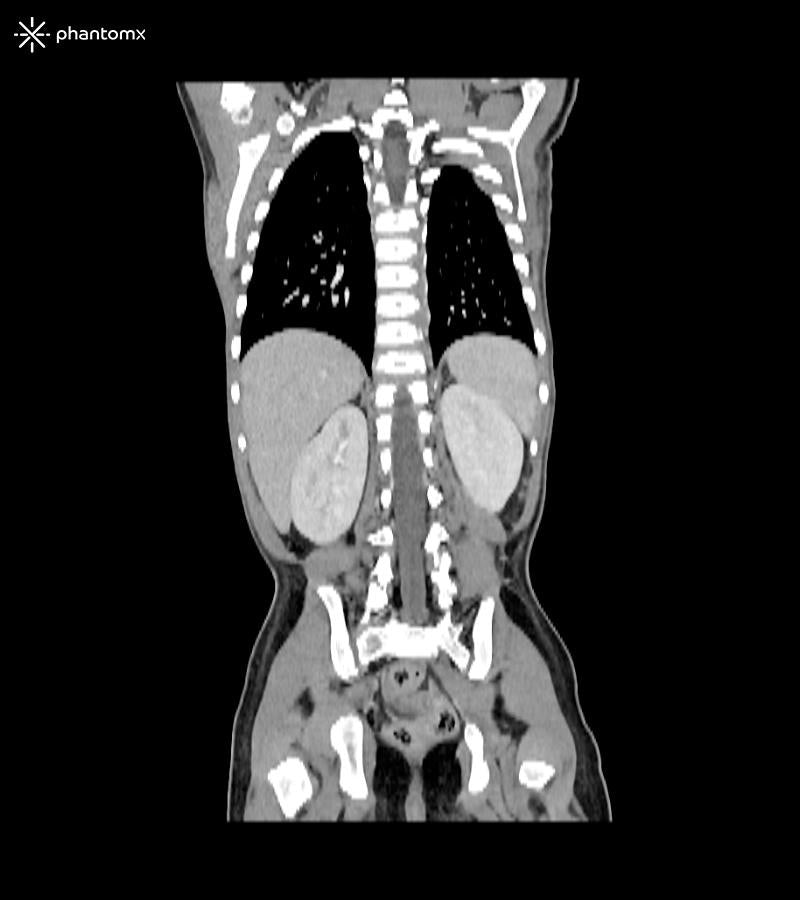
The Anatomy Phantom Models for CT, X-Ray, and Radiation Therapy accurately replicate anatomical structures and tissue contrasts, providing a lifelike representation during imaging and radiation therapy procedures.
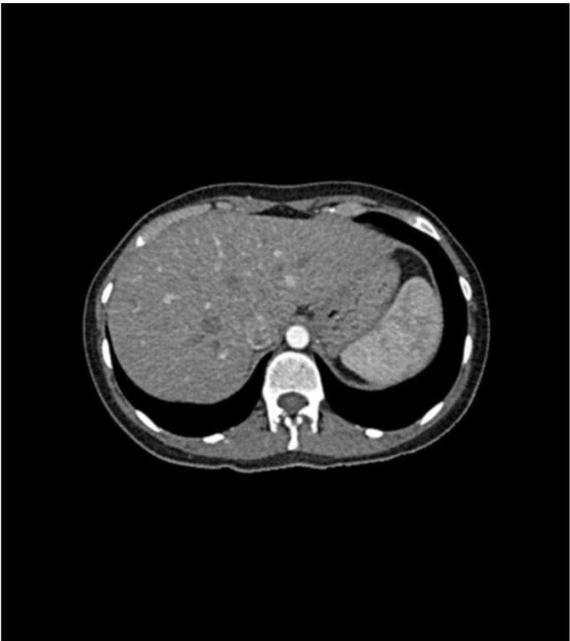
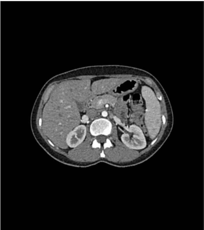
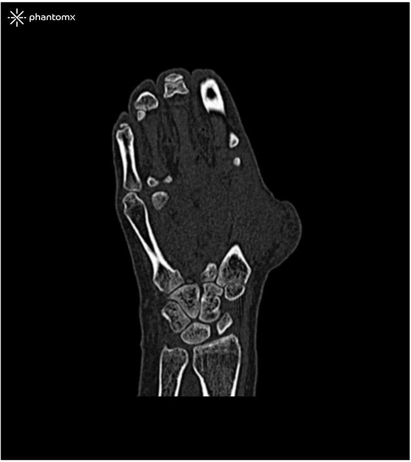
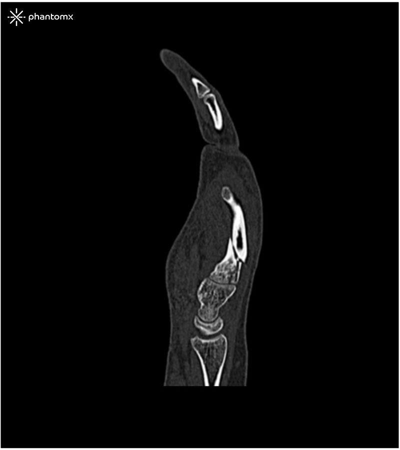
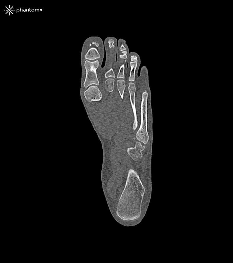
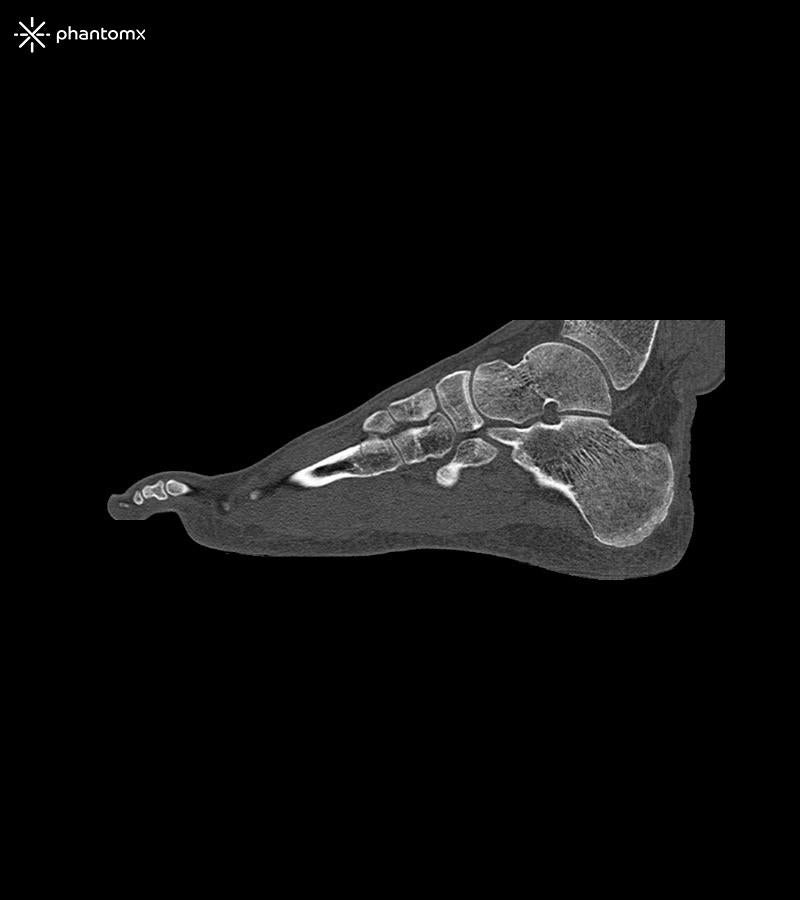
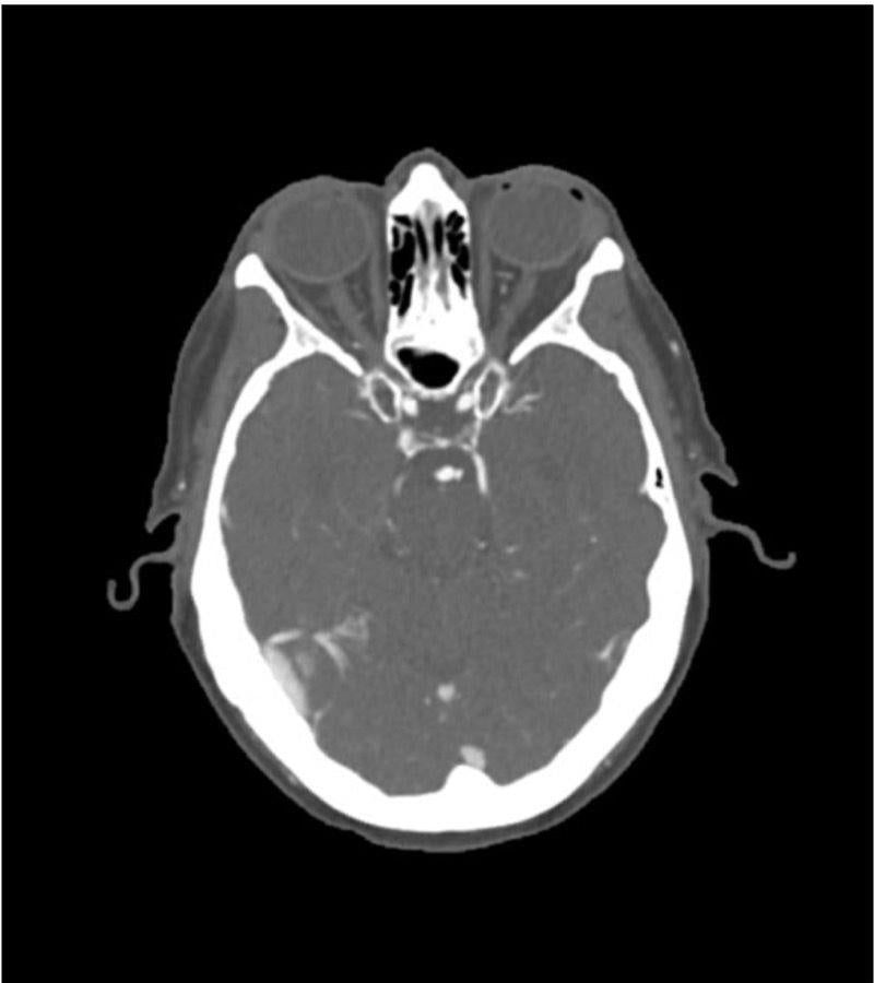
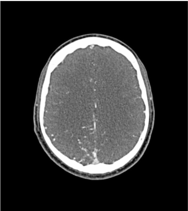
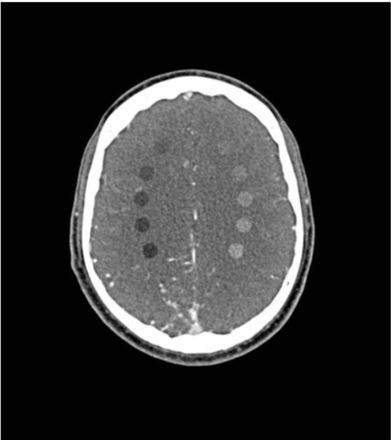
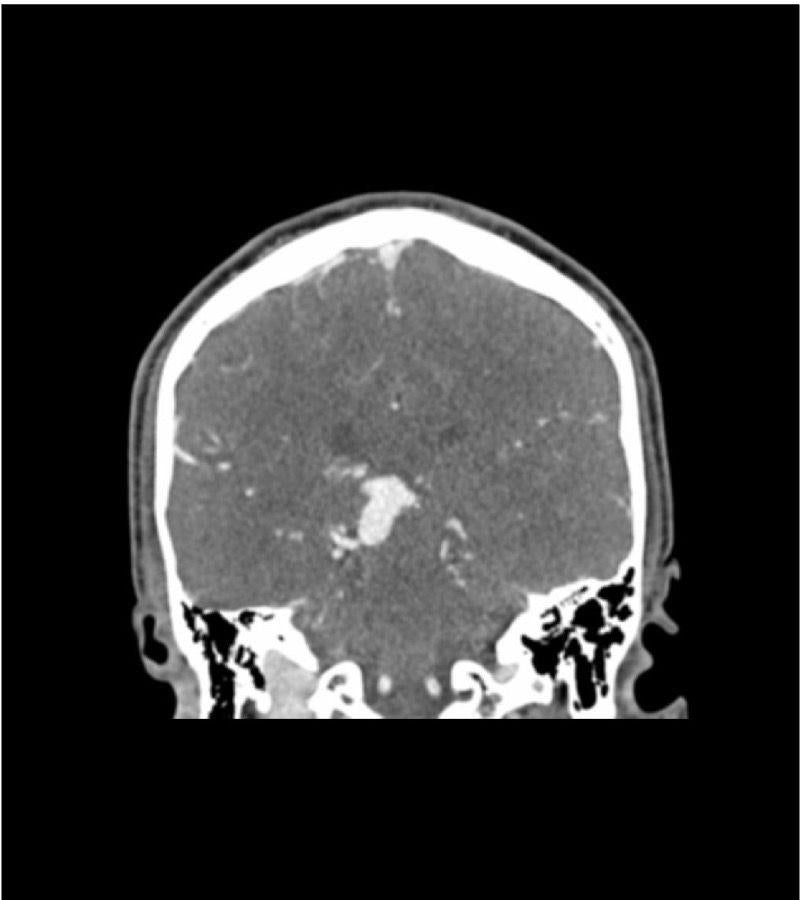
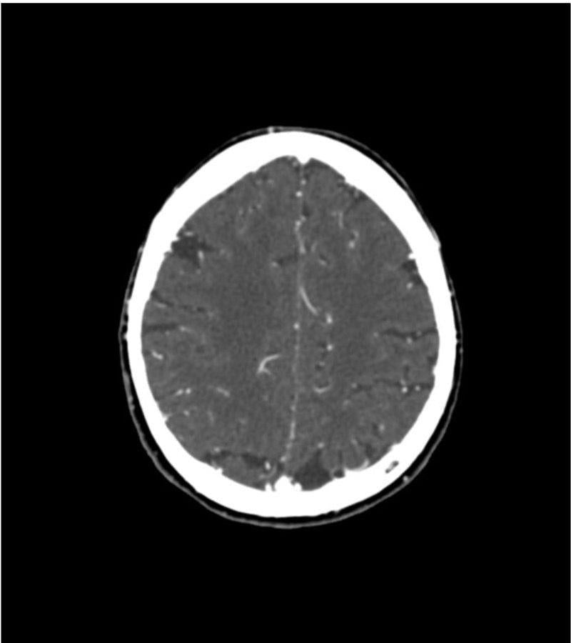
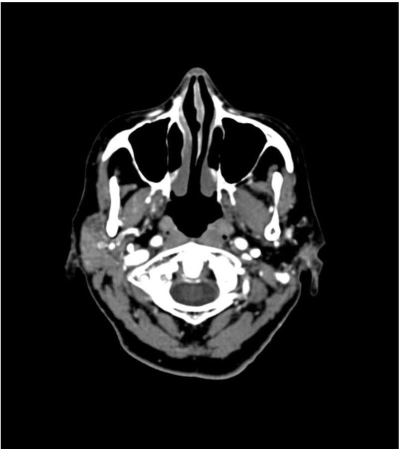
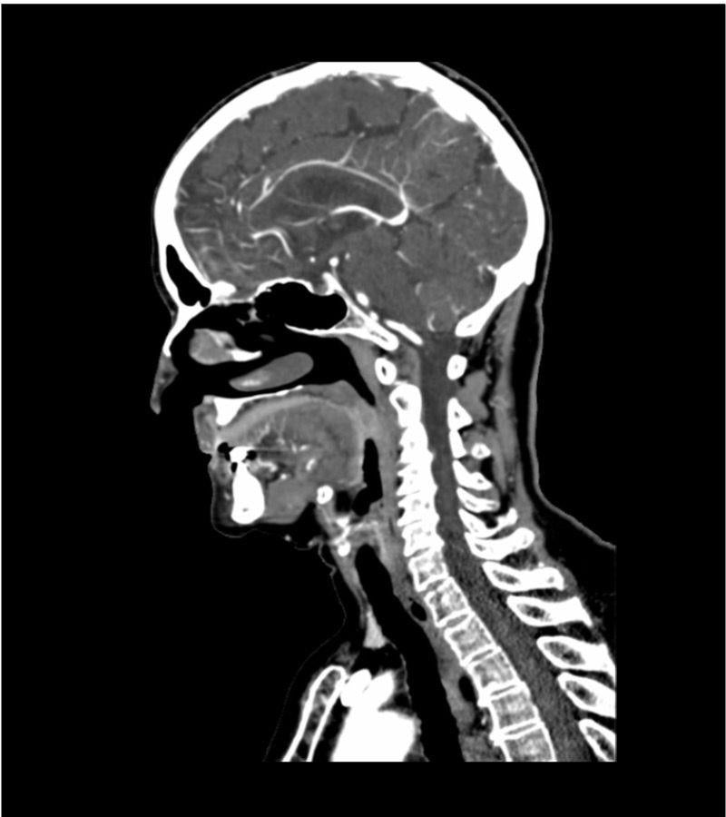
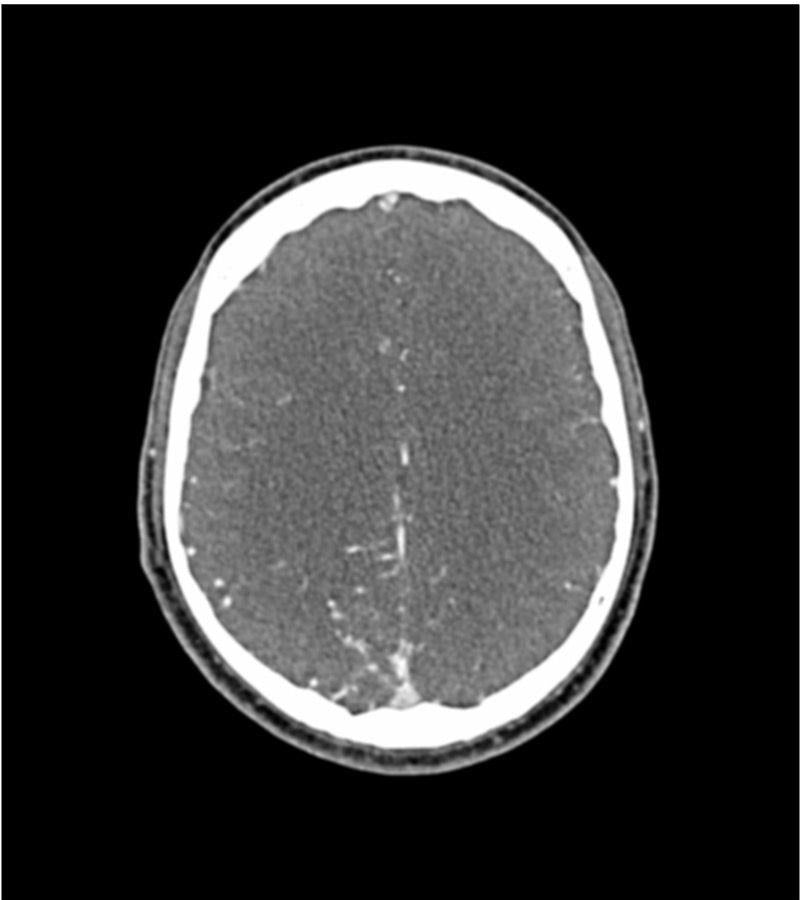
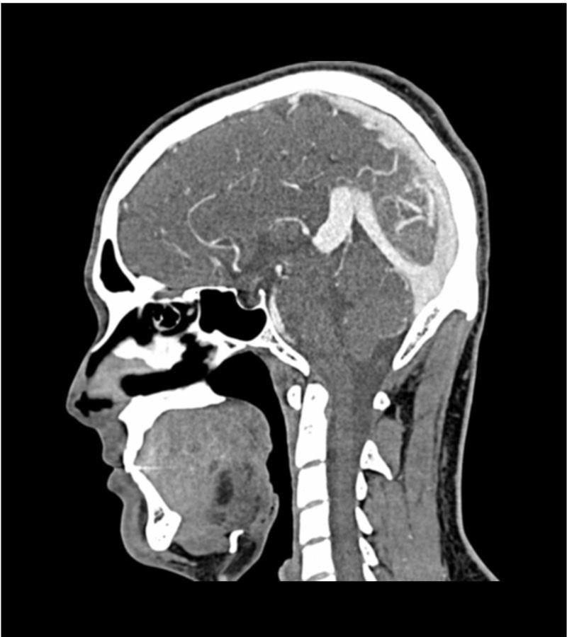
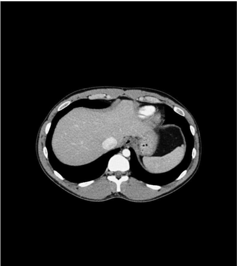
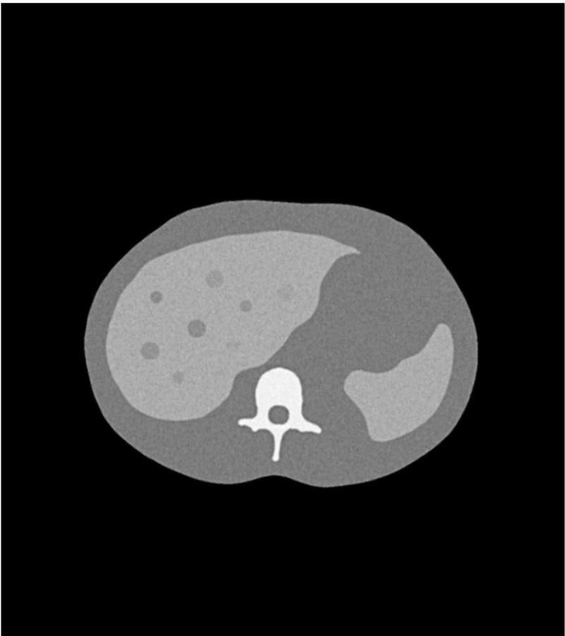
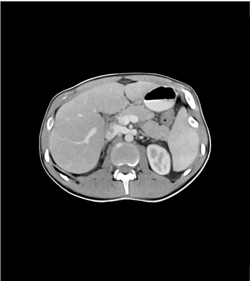
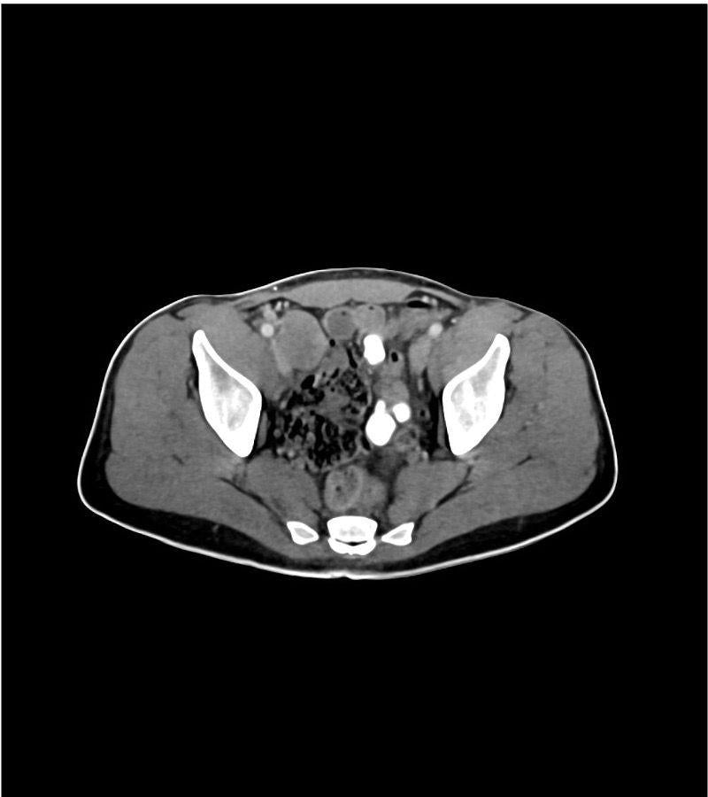
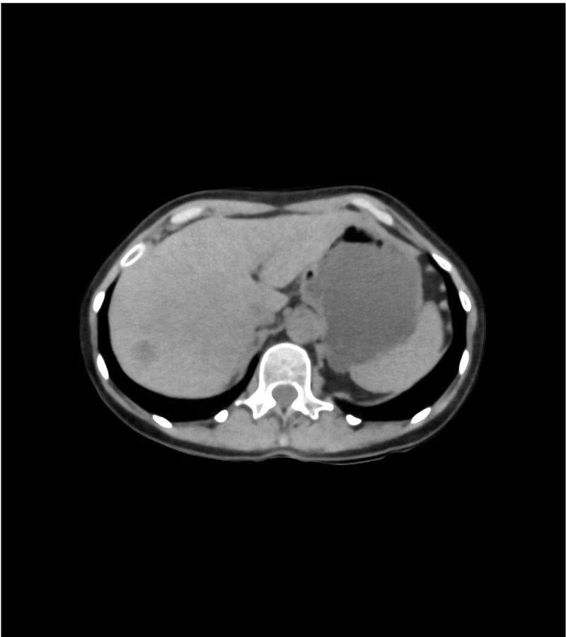
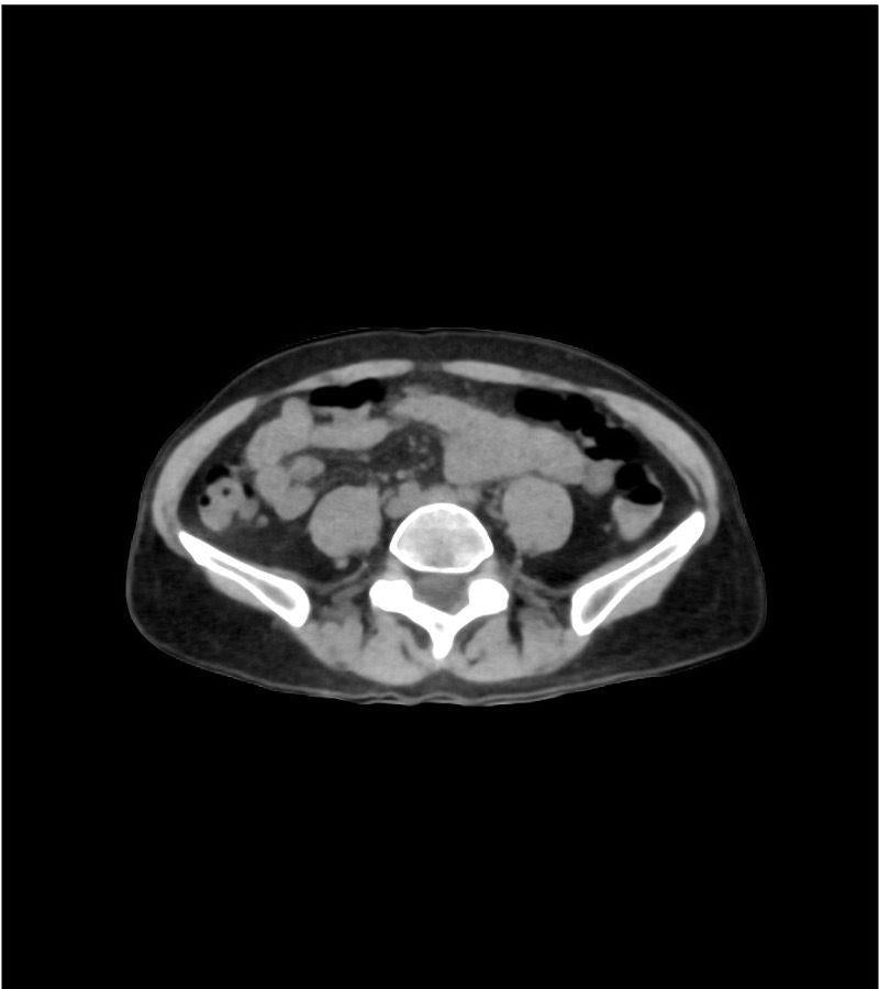
The anatomy phantom models were created using state-of-the-art technology and are based on actual patient data, ensuring authenticity. They accurately display bones, vessels, and soft tissue with realistic values at 120 kVp tube voltage in CT scans. If used with different tube voltages, such as 100 kVp, the CT values can be adjusted for proper calibration.
The phantom models exhibit realistic tissue contrasts in X-ray imaging, and air spaces are filled with a material measuring approximately -80 Hounsfield units.