


Abdominal Aortic Aneurysm
The specimen features a 10cm x 7cm abdominal aortic aneurysm below the renal arteries, with chronicity indicated by a laminated thrombus. Recent thrombus evidence and aneurysmal dilatation of adjacent arteries are observed. Focal ulcerated atheromatous plaques are present in the upper abdominal aorta, with no signs of rupture.
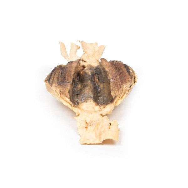
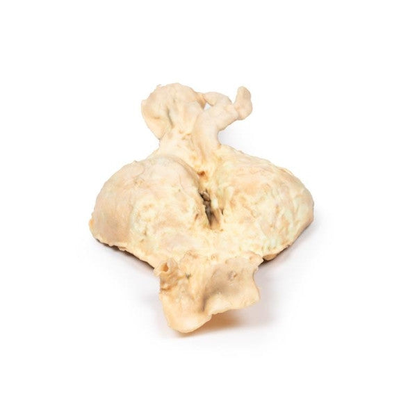
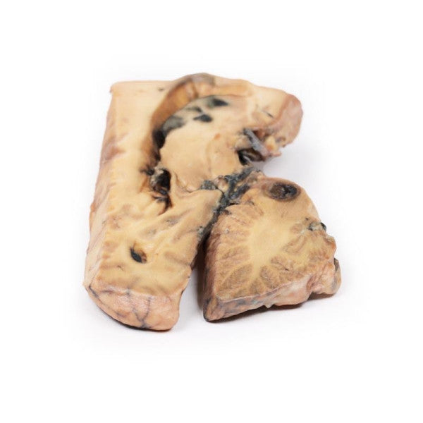
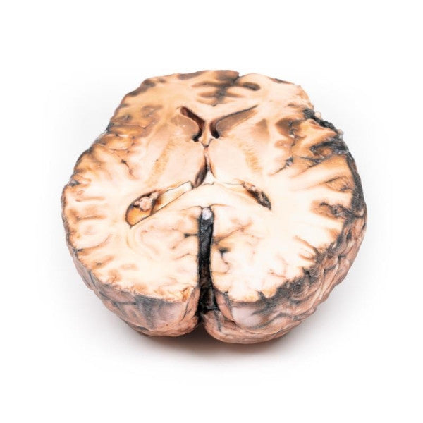
Berry Aneurysm of Basilar Artery
This midsagittal brain section exhibits a 5cm x 2cm basilar artery aneurysm extending into the midbrain and pons, filled with a thrombus. The lateral view reveals ventricle dilatation, blood staining, and haemorrhagic infarction of the caudate nucleus.
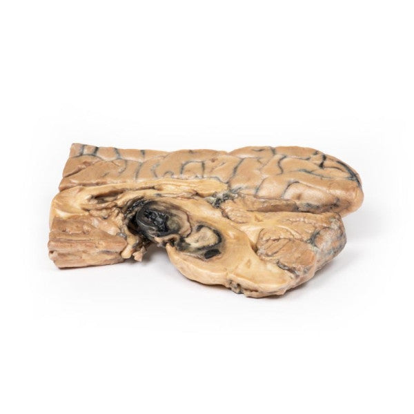
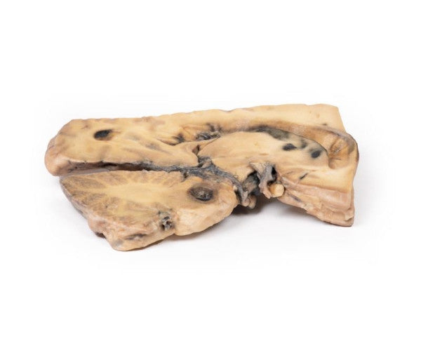
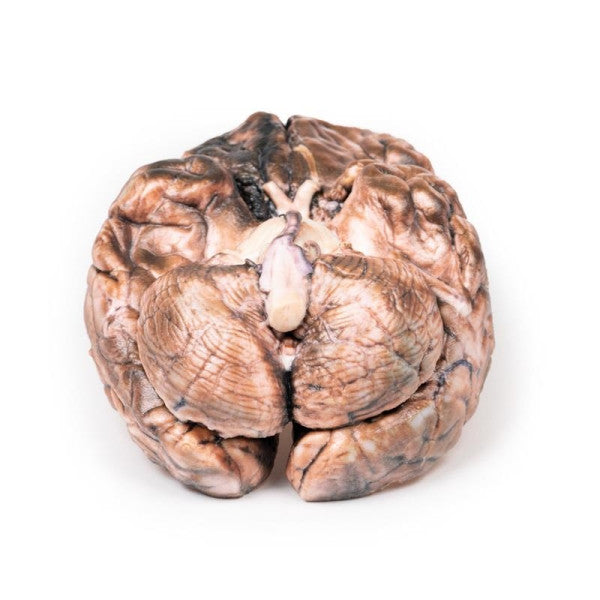
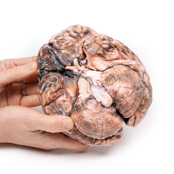
Ruptured Berry Aneurysm
The specimen reveals a ruptured 5mm saccular aneurysm at the right internal carotid and posterior communicating artery junction, with subarachnoid blood in the surrounding areas. An unruptured aneurysm is present on the left side, and the anterior portion of the right frontal lobe appears softer and more friable.
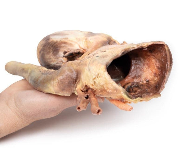
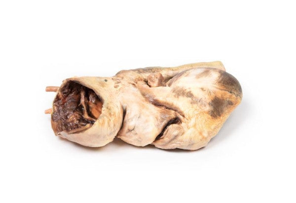
Ruptured Thoracic Aortic Aneurysm
The heart exhibits the posterior view of both ventricles. The thoracic ascending aorta reveals a significant saccular dilatation with visible atherosclerotic plaques and evidence of rupture, indicated by dark staining. Both ventricles display hypertrophy, while the coronary arteries, aortic valve, and tricuspid valve appear normal.
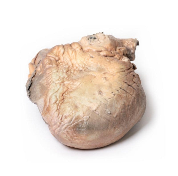
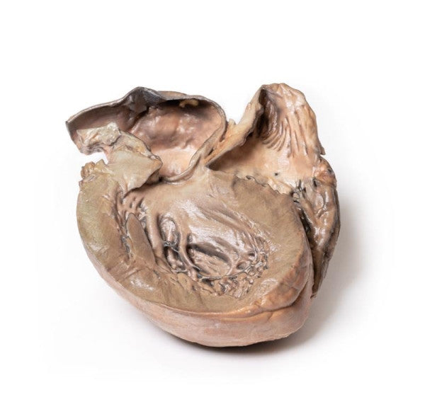
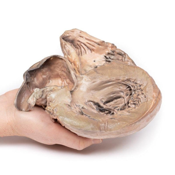
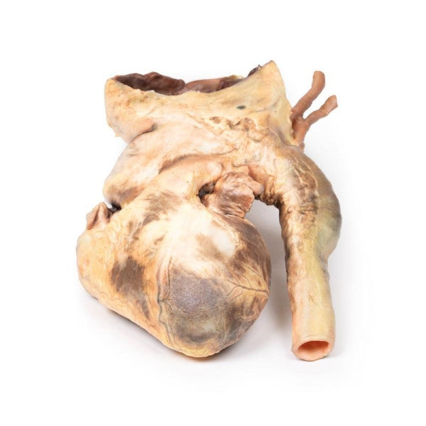
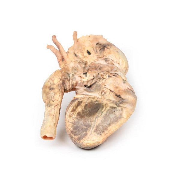
Syphilitic Aneurysm
This enlarged heart, with a dilated ascending aorta and a large aneurysm, shows scarred surfaces and atheroma. Arteries are displaced, and a ridge on the aneurysm's internal surface corresponds to pericardial sac attachment. Marked blood vessel congestion indicates syphilitic involvement.