


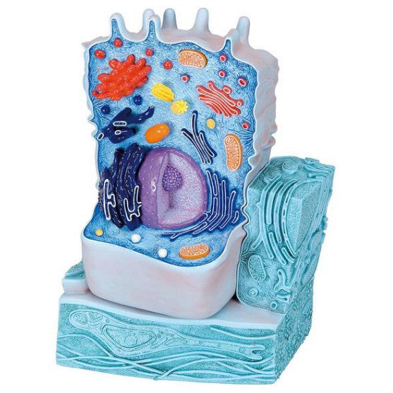
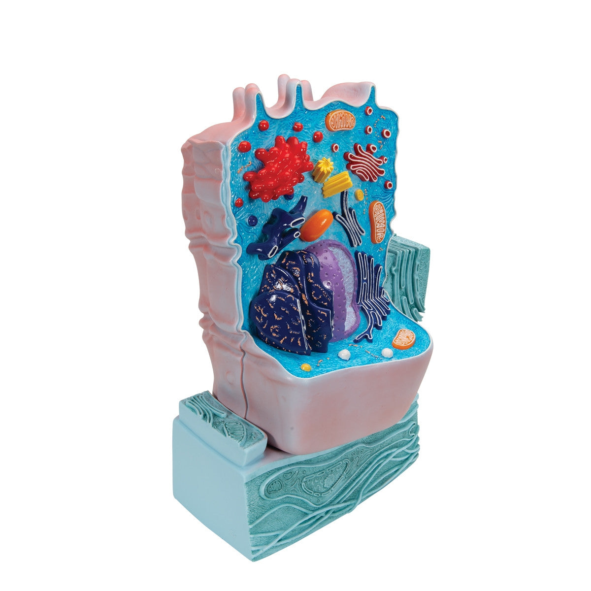
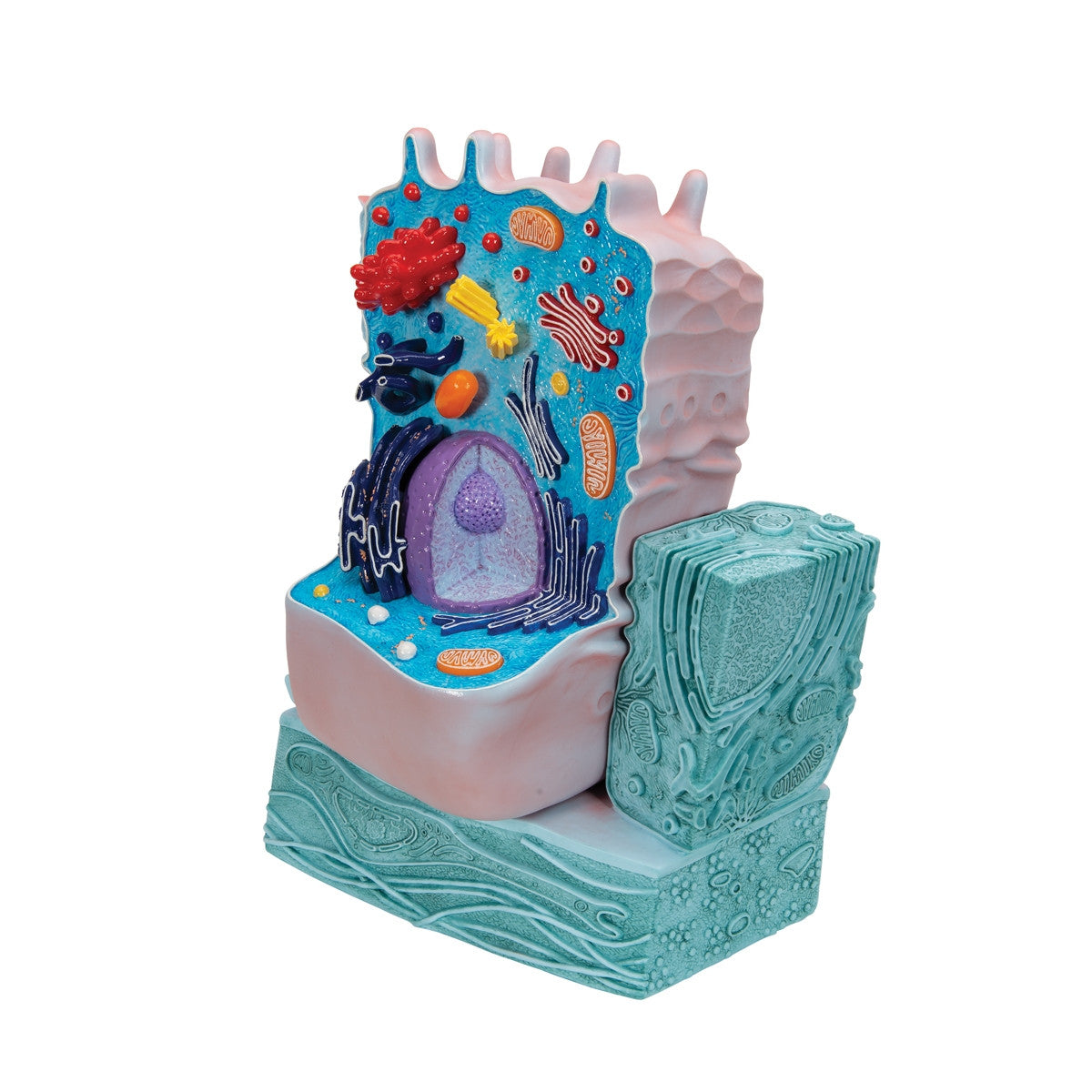
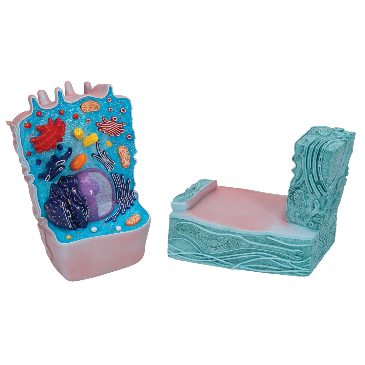
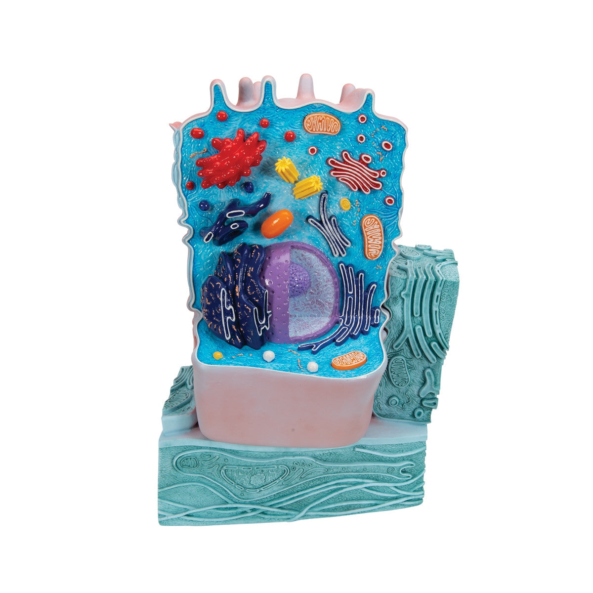
This educational model represents the structure and form of a typical animal cell when viewed by an electron microscope. To enable better study, the important organelles are coloured and in raised relief. The structures featured in the animal cell model include lysosome, microvilli, golgi apparatus, collagen fibres, basal membrane, rough and smooth endoplasmic reticulum, mitochondrion and nucleus. This model R04 / 1000523 would be an ideal teaching aid in a science classroom or lab.
This model comes with 3B Scientific 3B Smart Anatomy app included for FREE. This features access to an anatomy course, including 3 digital anatomy lectures, 117 different virtual anatomy models and 39 anatomy quizzes. It also offers a FREE warranty upgrade from 3 to 5 years with every product registration. To unlock these benefits, scan the label located on your 3B Scientific anatomy model and register online.
Part sh
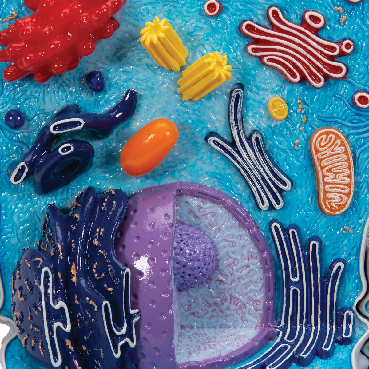
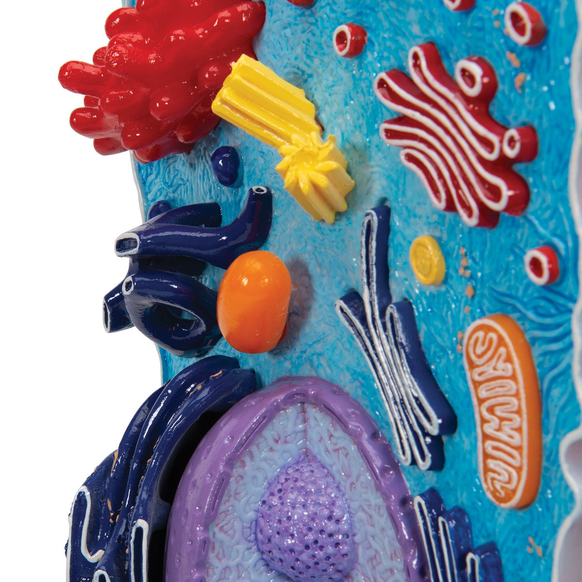
1 Cell nucleus
2 Nucleolus
3 Mitochondrion
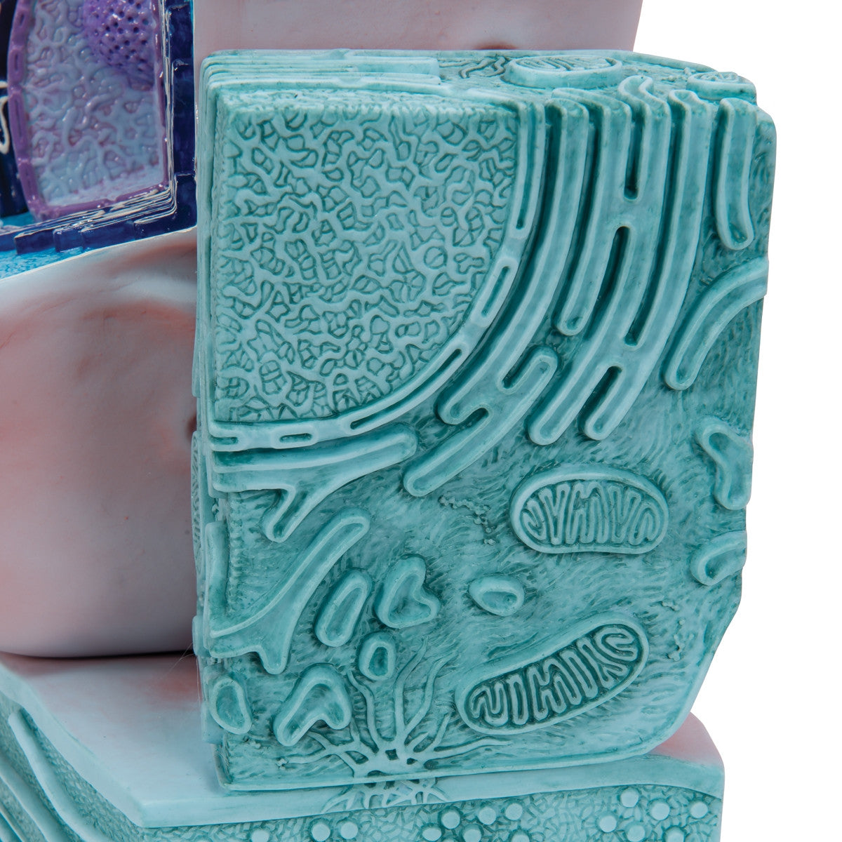
4 Smooth endoplasmic reticulum (ER)
5 Desmosome (Macula adhaerens)
6 Basal membrane
7 Hemidesmosome
8 Collagen fibres
9 Fibroblast
10 Peroxisome
11 Lysosome
12 Rough endoplasmic reticulum (ER)
13 Mitochondrion
14 Smooth endoplasmic reticulum (ER)
15 Golgi Apparatus
16 Centriole
17 Cytosol with embedded filament of the cytoskeleton
18 Microvilli
19 Secretion vesicle
20 Golgi Apparatus
21 Lysosome
22 Zonula occludens
23 Zonula adhaerens
24 Desmosome (Macula adhaerens)
25 Microplica