


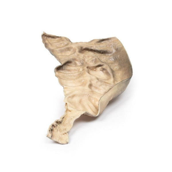
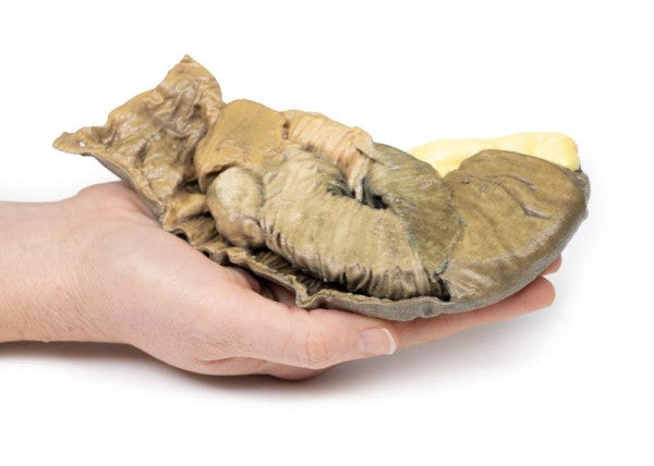
Hirschsprung's Disease
This postmortem sigmoid colon section reveals a significantly dilated proximal bowel (sigmoid) with the loss of the usual mucosal pattern. The distal bowel (rectum) has a regular diameter but lacks ganglion cells in the myenteric plexus, illustrating Hirschsprung's disease or congenital aganglionic megacolon.
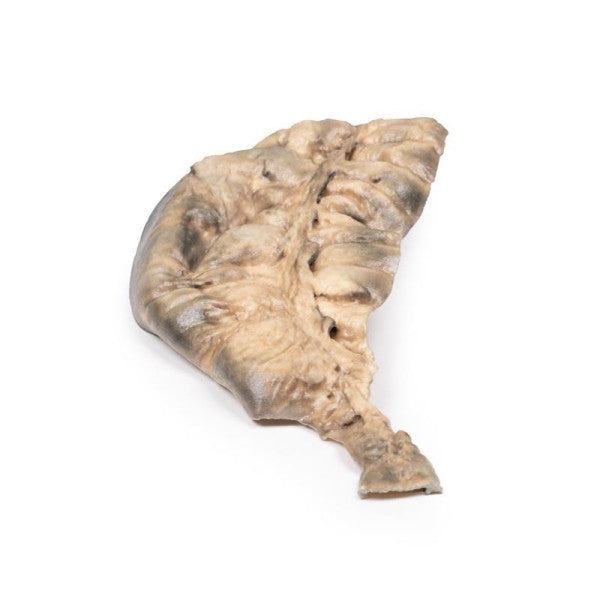
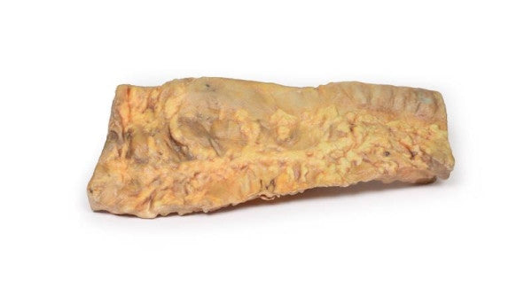
Intussusception of Small Bowel Due to Metastatic Tumour
This bowel specimen includes a 20 cm segment with a 2 cm wide attached mesentery. A 3 cm polypoid tumour near the proximal margin has caused a 13 cm intussusception, showing signs of early ischaemic necrosis. The tumour, located at the intussusception apex, appears macroscopically consistent with a metastatic malignant tumour, with the potential for a primary tumour.
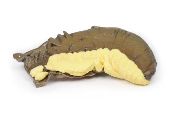
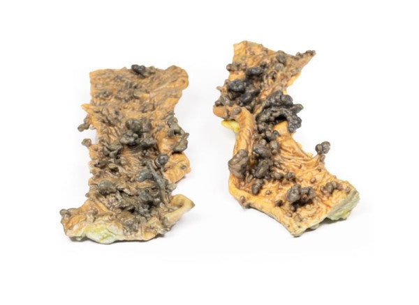
Multiple Polyposis Coli
Two segments of the sigmoid colon are presented. The bowel's mucosa is adorned with multiple partially pigmented polyps, both sessile and pedunculated, with a maximum diameter of 1.5 cm. No macroscopic signs of malignancy observed.
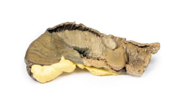
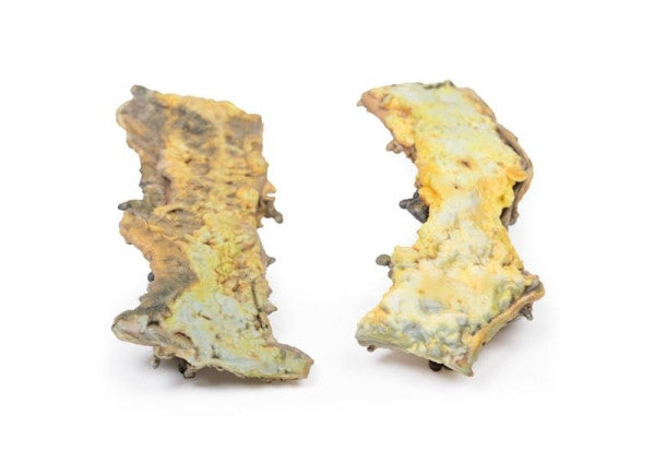
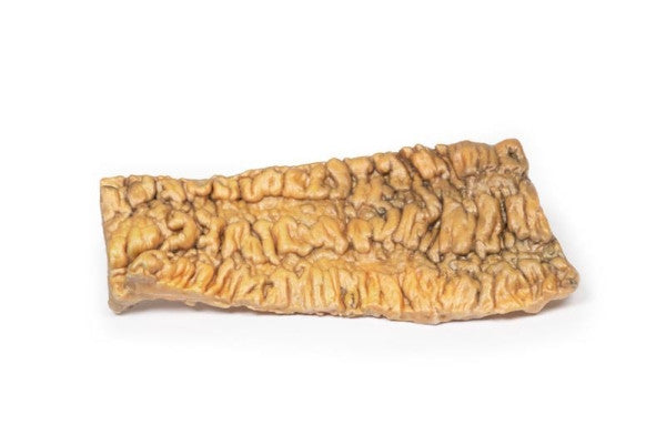
Ulcerative Colitis
The excised colon reveals widespread ulceration with oedematous patches of remaining mucosa. The ulcers exhibit necrotic bases, and some edges create "pseudo"-polyps. Histological examination of the bowel mucosa indicates acute inflammatory changes, crypt abscesses, focal necrosis, and ulceration, characteristic of acute ulcerative colitis.