


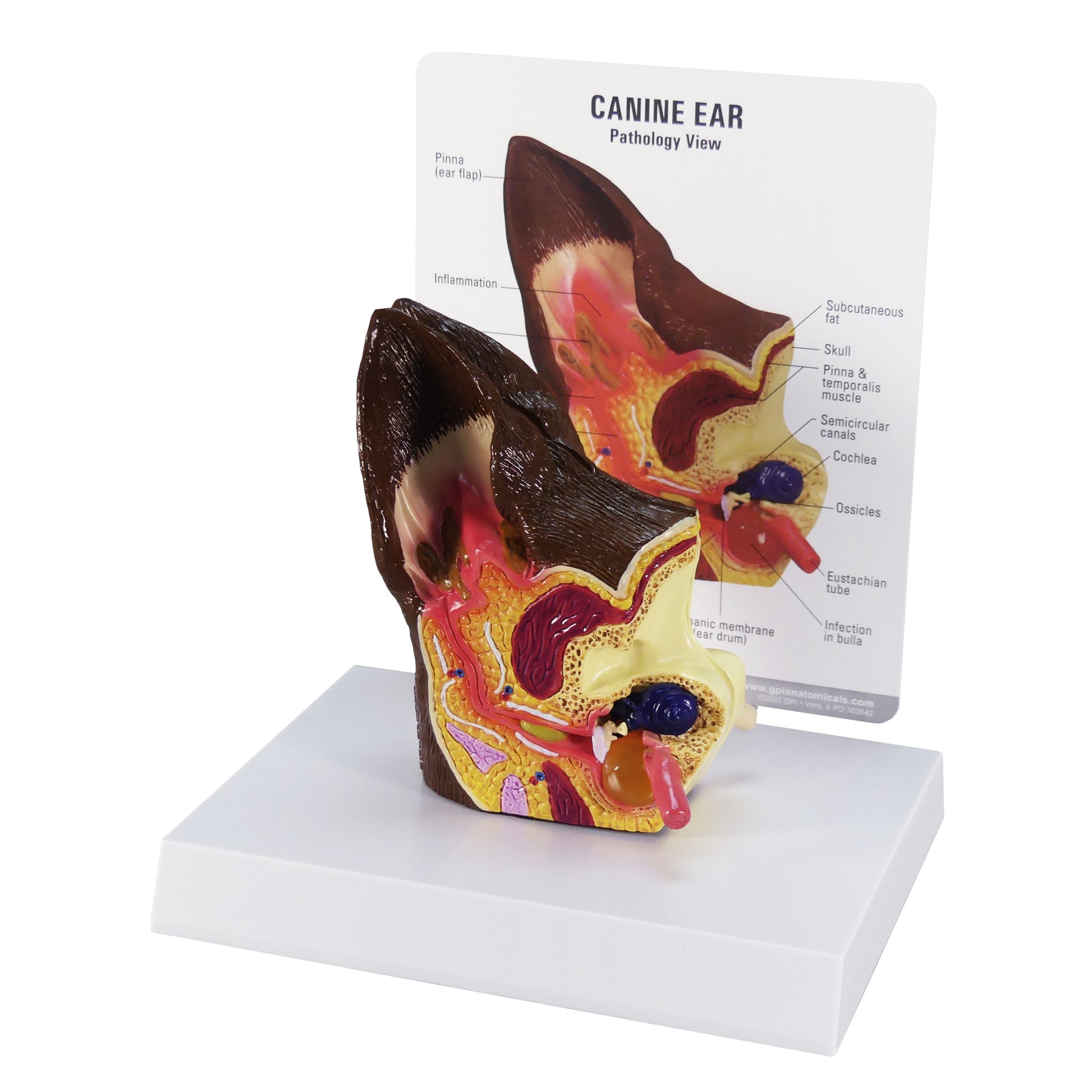
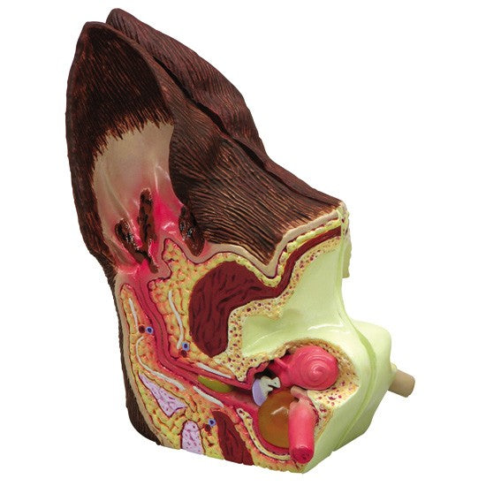
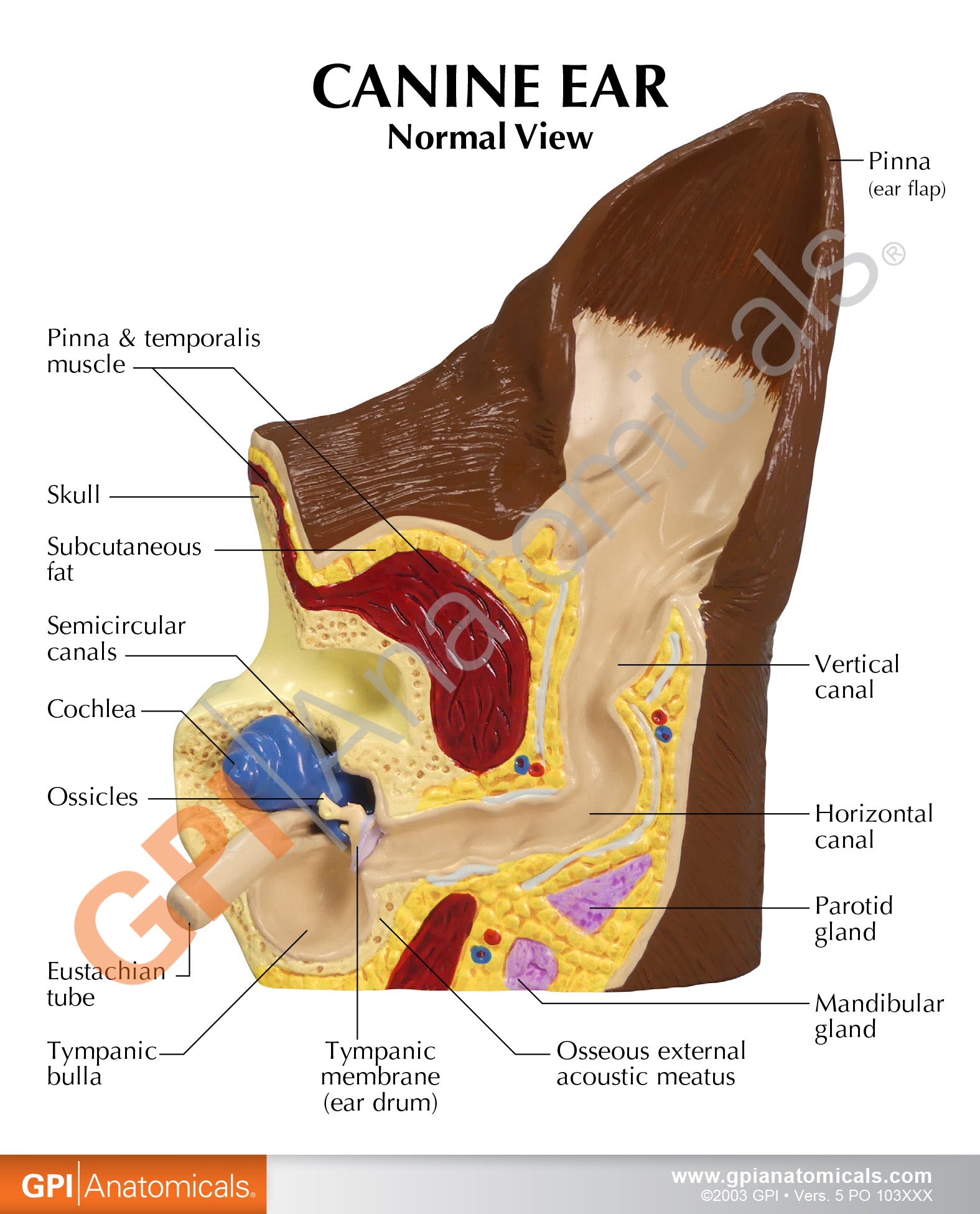
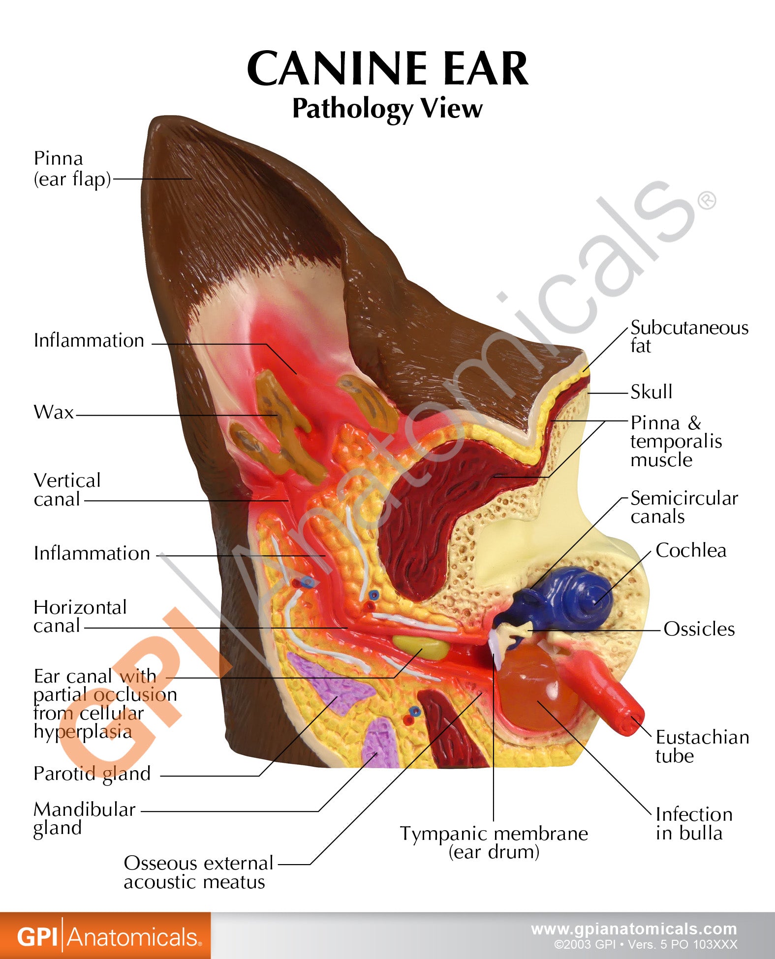
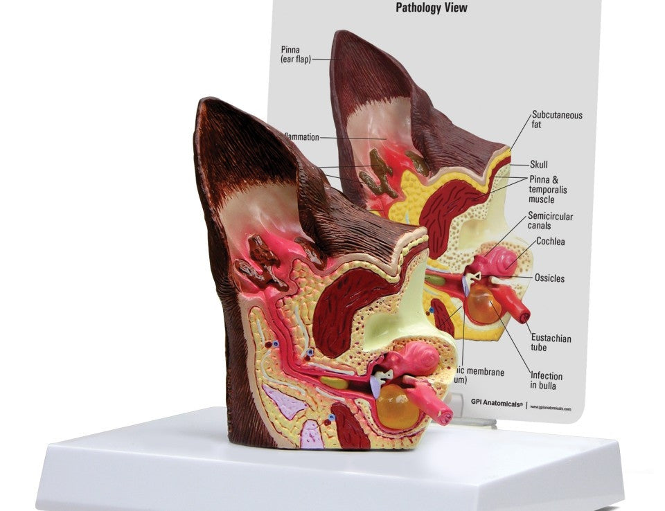
This life-Size Canine Ear Model depicts both healthy and diseased anatomy. The normal side shows the cochlea, auditory ossicles, auditory tube, tympanic bulla, middle ear cavity, tympanic membrane, horizontal canal, vertical canal, auricular cartilage, pinna and temporalis muscle; the diseased side illustrates inflamed inner ear structures, inflammatory exudate in tympanic bulla, ear canal with partial occlusion from cellular hyperplasia, inflammatory exudate and an inflamed (reddened) outer ear.
This ear model is ideal for demonstrating anatomy and diseases affecting dogs. The veterinary anatomy model comes with a two-sided education card included. An ideal tool for vets to explain dog anatomy to pet owners, and common conditions affecting their pets. NB: Model is based on the ear size of an average size dog.
AnatomyStuff.co.uk is the exclusive UK distributor for the GPI Anatomicals vet anatomy model range. If you would like to find out more about adding your logo to these models for promotional purposes, please contact us for more information.