


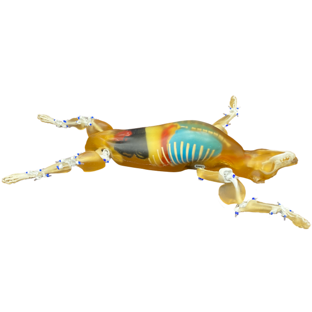
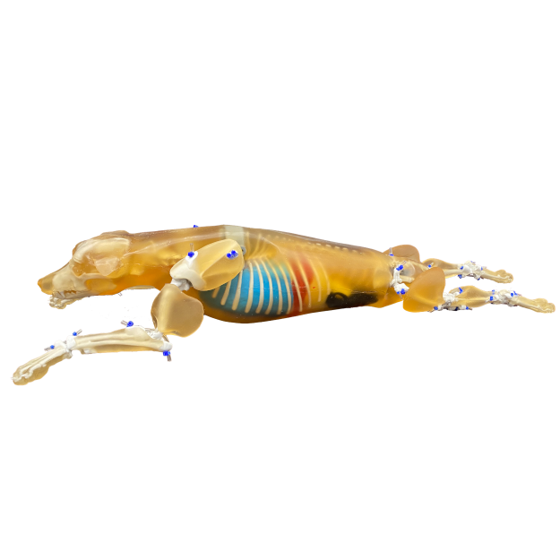
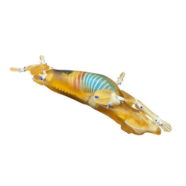
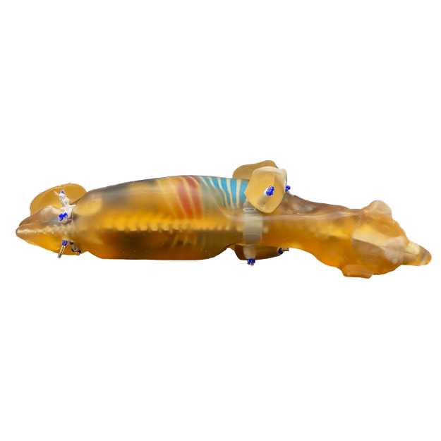
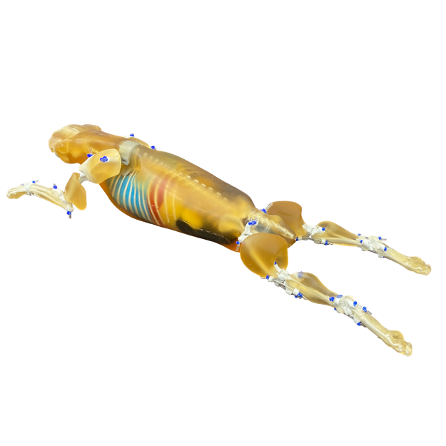
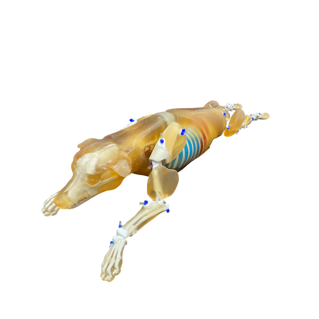
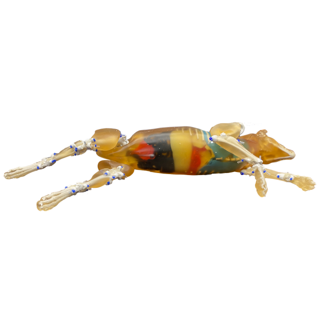
Introducing the Canine Phantom for X-Ray, CT, Ultrasound & MRI, this advanced Canine Phantom is designed for realistic veterinary imaging and clinical training across X-ray, CT, ultrasound and MRI. Developed from a real CT dataset of a 2-year-old male Bull Mastiff x Rhodesian Ridgeback and created using advanced AI-driven modelling and refined in collaboration with leading veterinary institutions in the UK, Europe, North America and Australia, it delivers exceptional anatomical accuracy for hands-on education and procedural practice.
Scaled to a manageable 16kg for ease of handling, the phantom includes both skeletal and soft-tissue structures, making it ideal for repeated use in academic and clinical environments.
Key Features
- Realistic joint articulation for lifelike limb positioning
- High anatomical accuracy based on real CT data
- Durable construction suitable for frequent training use
- Detailed vascular structures including liver, spleen and pancreas
- Fully modelled heart with chambers, valves and major vessels
- Complete gastrointestinal tract including small and large intestines
Imaging Compatibility
- Optimised for T2-weighted MRI with realistic tissue relaxation values
- Excellent performance in proton-density imaging
- Compatible with T1-weighted MRI, CT, X-ray and ultrasound
Included Anatomy
- Adult canine head, skull and jaw
- Complete spine, ribcage, pelvis and shoulders
- Realistic heart with major vessels (IVC, SVC, aorta)
- Lungs, diaphragm and abdominal organs
- Liver, kidneys, spleen and pancreas with vasculature
- Stomach, small and large intestines, caecum and rectum
- Bladder and ureters
- Front and rear legs
Construction & Contents
- Soft tissues and organs: urethane-based soft resin
- Bones: 3D-printed synthetic bone
- Hard carry case
- User manual and assembly instructions
Custom organ configurations are available on request.