


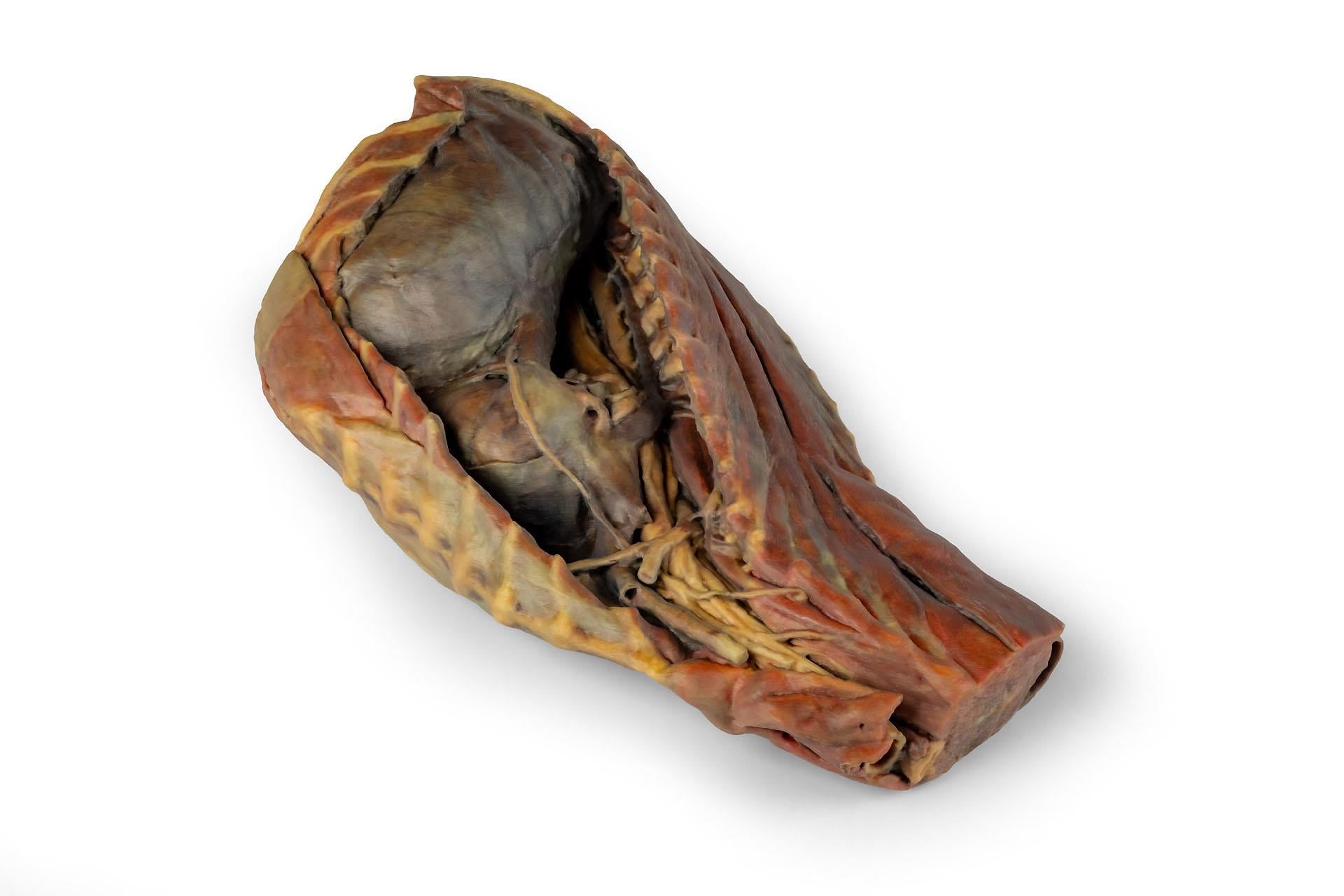
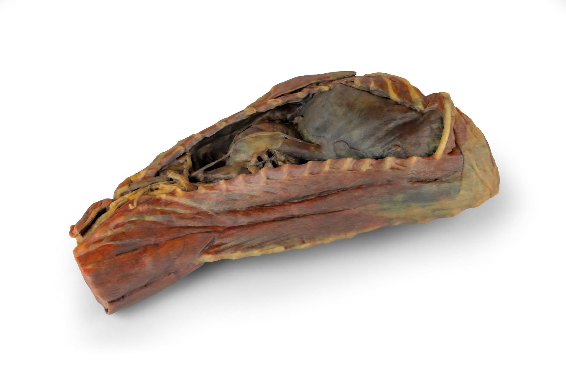
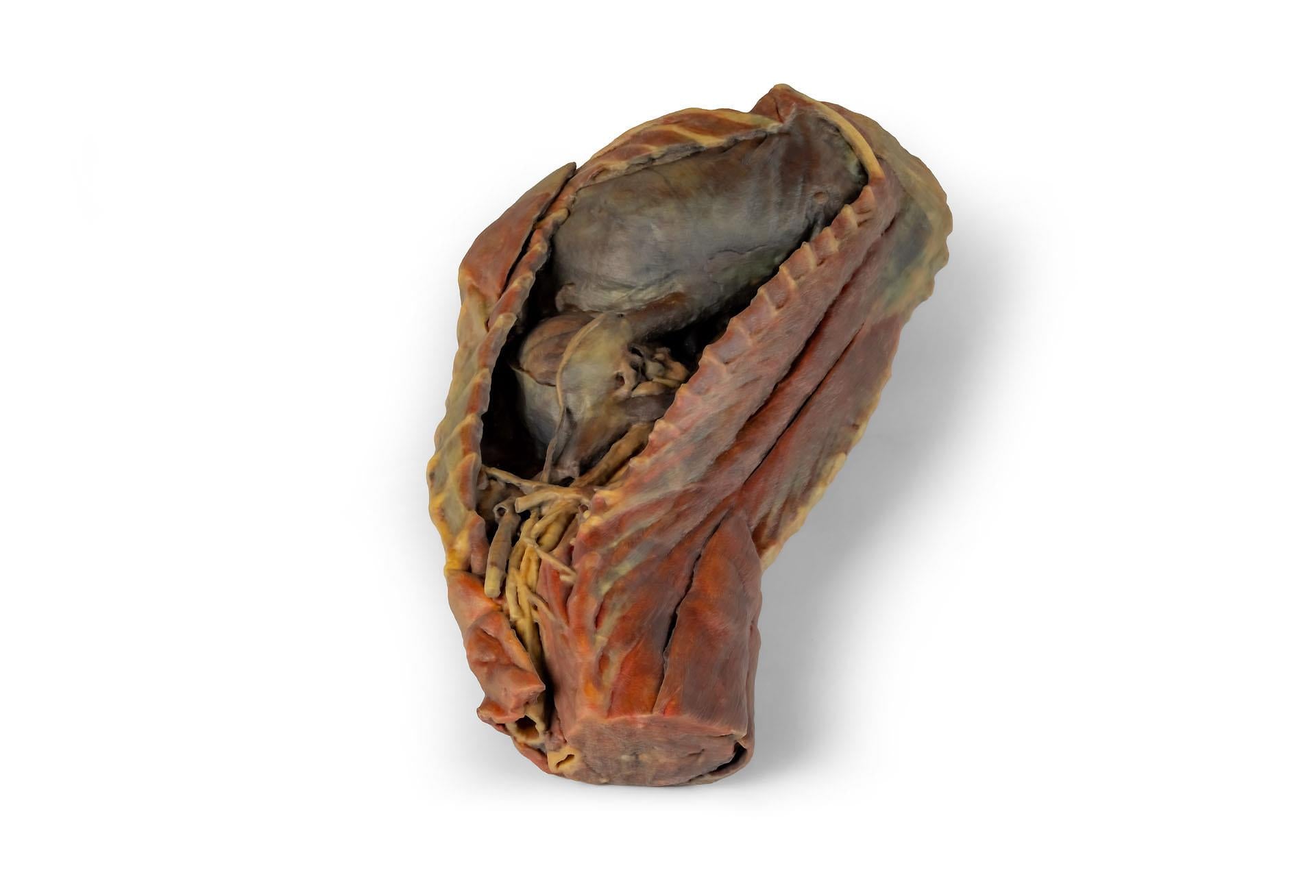
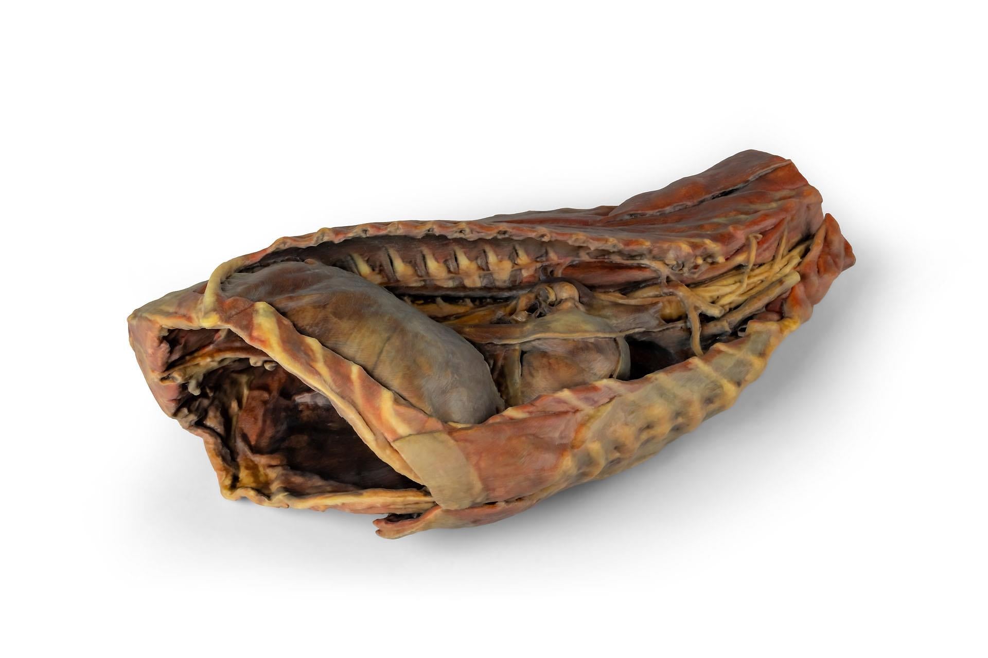
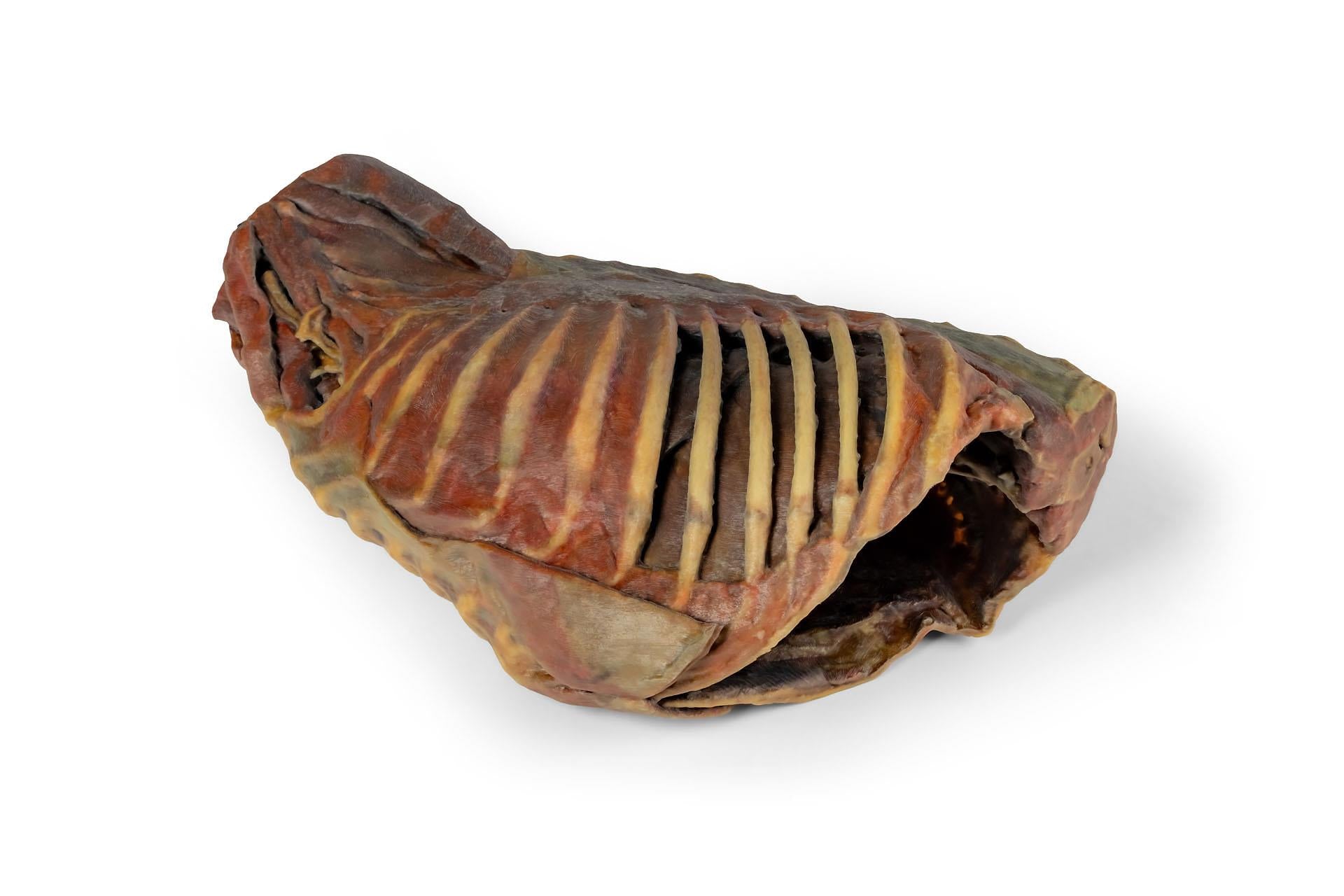
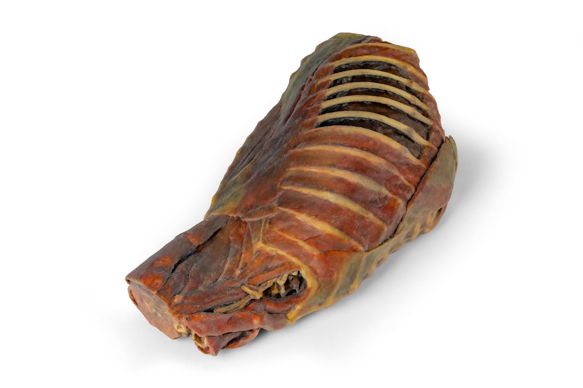
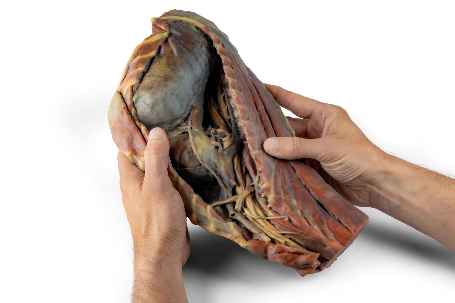
Enhance veterinary anatomy education with the highly detailed Canine Thoracic Cavity. This 3D-printed anatomical specimen presents a right-side dissection of the thoracic cavity of an adult dog, providing a comprehensive view of the thoracic organs, major vessels, nerves, and surrounding anatomical structures for advanced veterinary teaching and clinical study.
Following removal of the right thoracic wall and ribs, the right lung has been excised while preserving the pulmonary vessels in their natural anatomical relationship to the heart. The specimen clearly demonstrates the organisation of the cranial, middle, and caudal mediastinum, allowing detailed study of thoracic anatomy and associated structures.
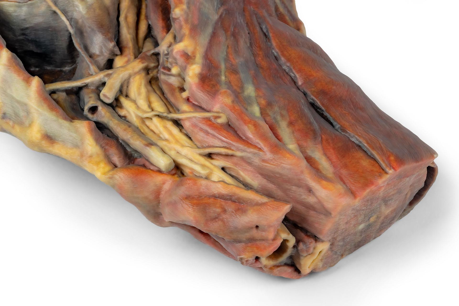
Visible structures within the cranial mediastinum include:
- Formation of the cranial vena cava
- Branches of the right subclavian artery
- Thoracic trachea and thoracic oesophagus
- Thoracic portion of the longus colli muscle
- Phrenic nerve and vagosympathetic trunk
- Stellate ganglion
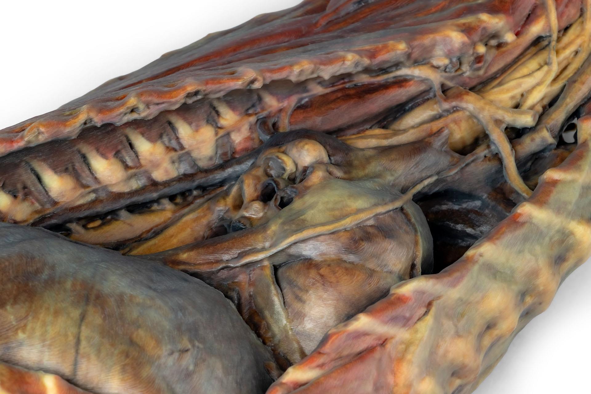
The middle mediastinum displays the heart partially enclosed by the fibrous pericardium, together with the pulmonary vessels and principal bronchi formed by division of the trachea.
Within the caudal mediastinum, the specimen demonstrates the course of the caudal vena cava, right phrenic nerve, thoracic aorta, right azygos vein, sympathetic trunk, and the dorsal and ventral vagal trunks associated with the oesophagus.
The preserved diaphragm clearly shows its attachments to the costal arches and lumbar vertebrae. Additional retained structures include the intercostal muscles, brachial plexus, and extensor musculature of the cervical and thoracic vertebral column.
The realistic dissection and accurate anatomical presentation make this specimen highly suitable for veterinary anatomy laboratories, pathology teaching, and advanced thoracic anatomy education.
Key features:
- 3D-printed from a real canine thoracic cavity specimen
- Detailed right-side thoracic cavity dissection
- Displays heart, mediastinum, major vessels, nerves, and bronchi
- Visible cranial, middle, and caudal mediastinal structures
- Preserved diaphragm, intercostal muscles, and brachial plexus
- Ideal for advanced veterinary anatomy and pathology education
Ideal for veterinary schools and universities for anatomy and pathology instruction as well as clinical and surgical training.