


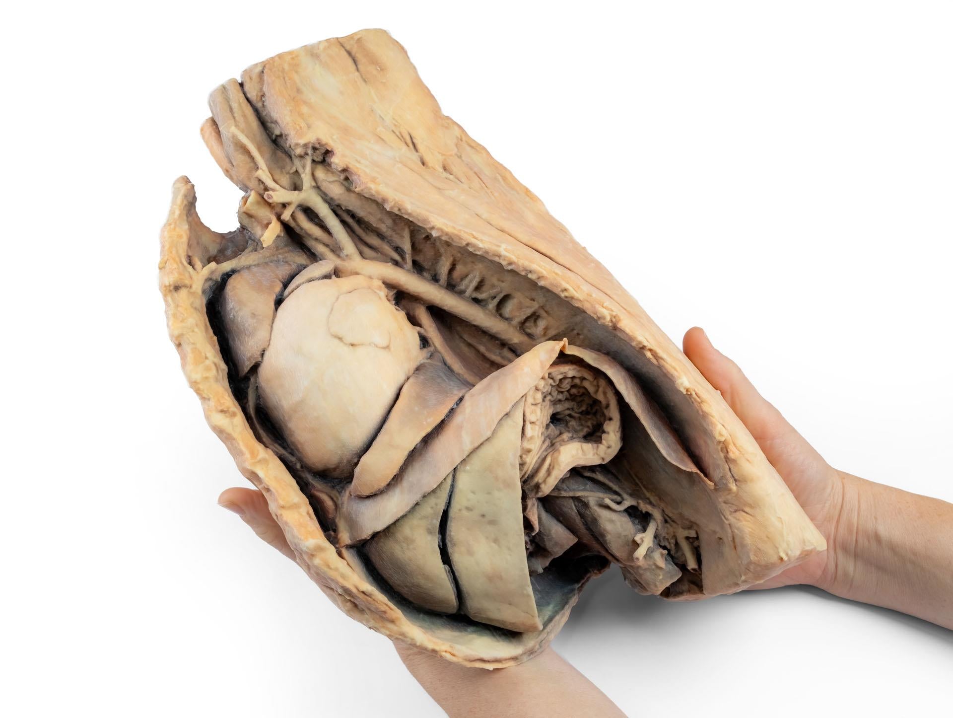
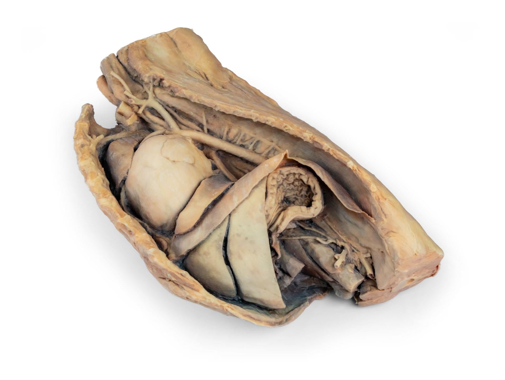
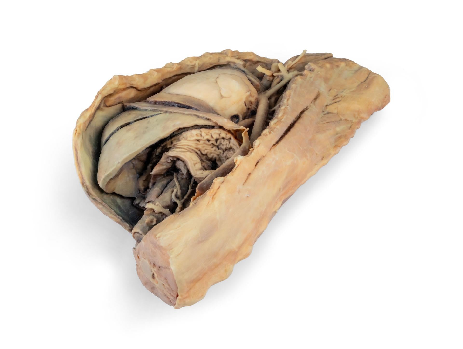
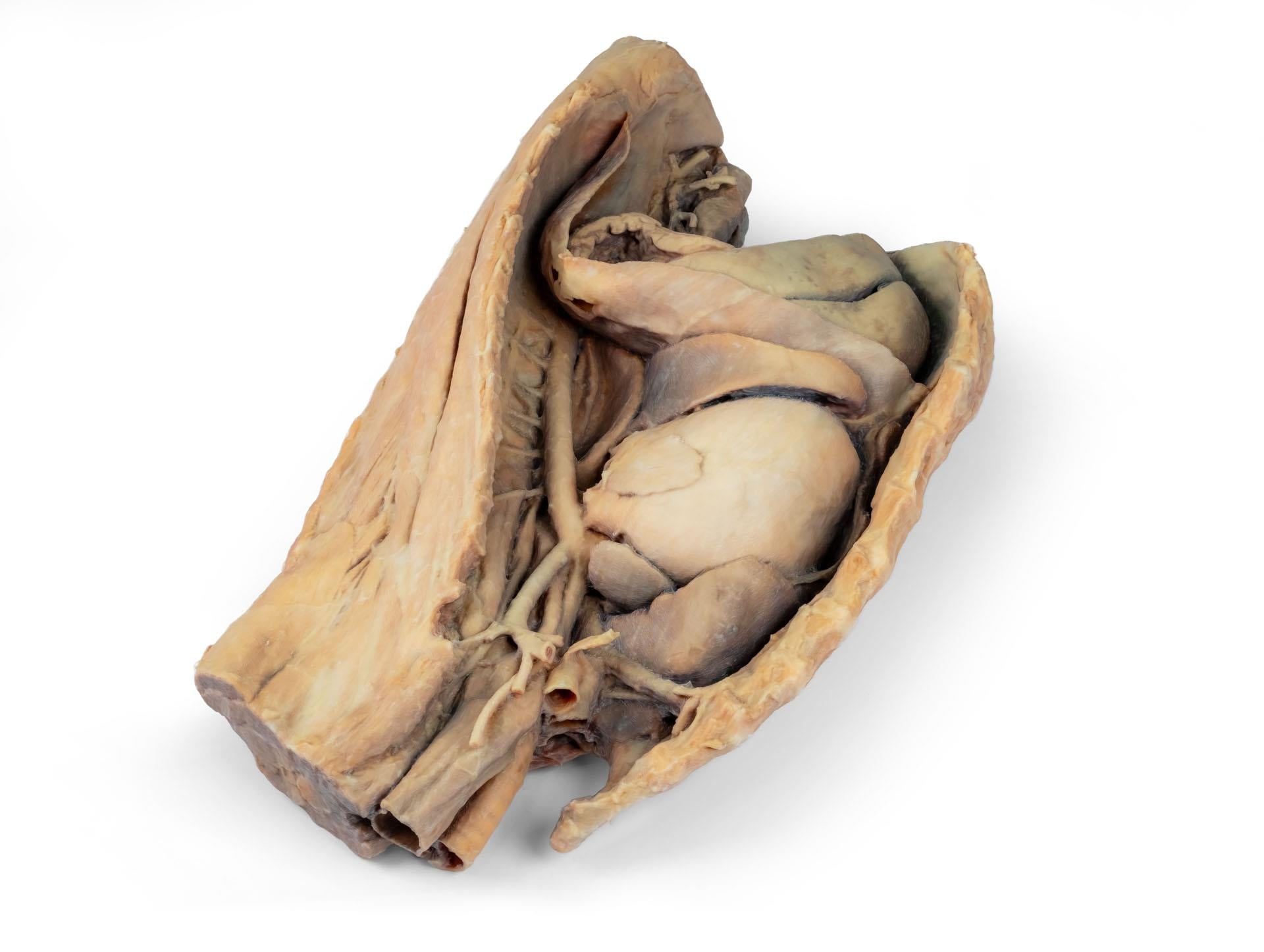
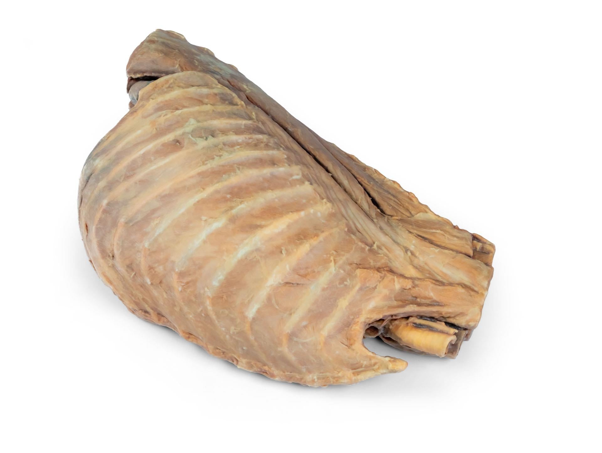
Enhance veterinary anatomy education with the highly detailed Canine Thoracic Cavity (Left Side). This 3D-printed anatomical specimen illustrates the topography of the heart following removal of the left lung during a left-sided dissection of the thoracic cavity and cranial abdomen in an adult dog, providing an exceptional teaching model for advanced anatomical study.
The cranial and accessory lobes of the right lung remain in situ, serving as important anatomical reference points for orientation within the thoracic cavity. The specimen clearly demonstrates the spatial relationships between the heart, great vessels, diaphragm, and upper abdominal structures.
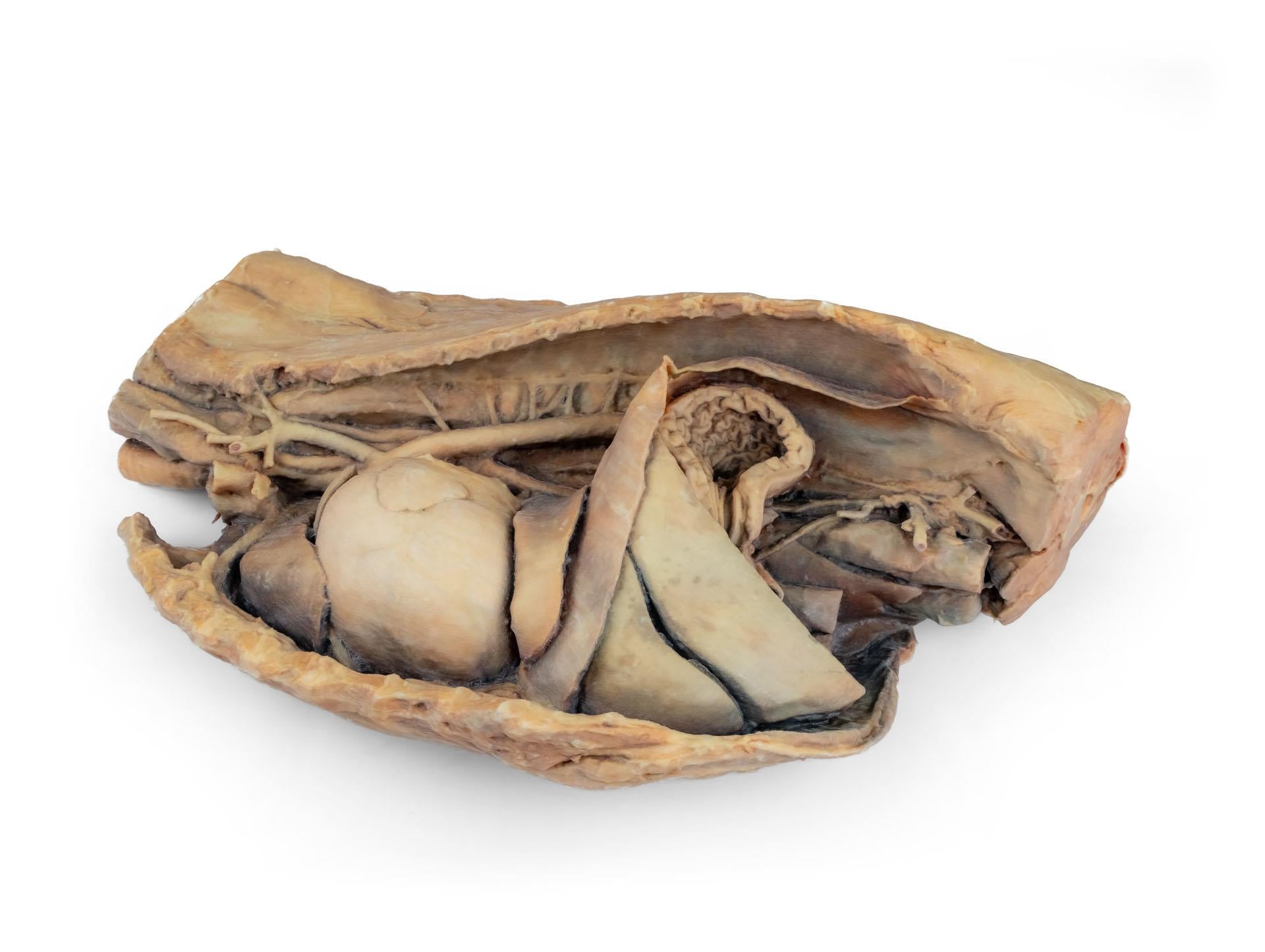
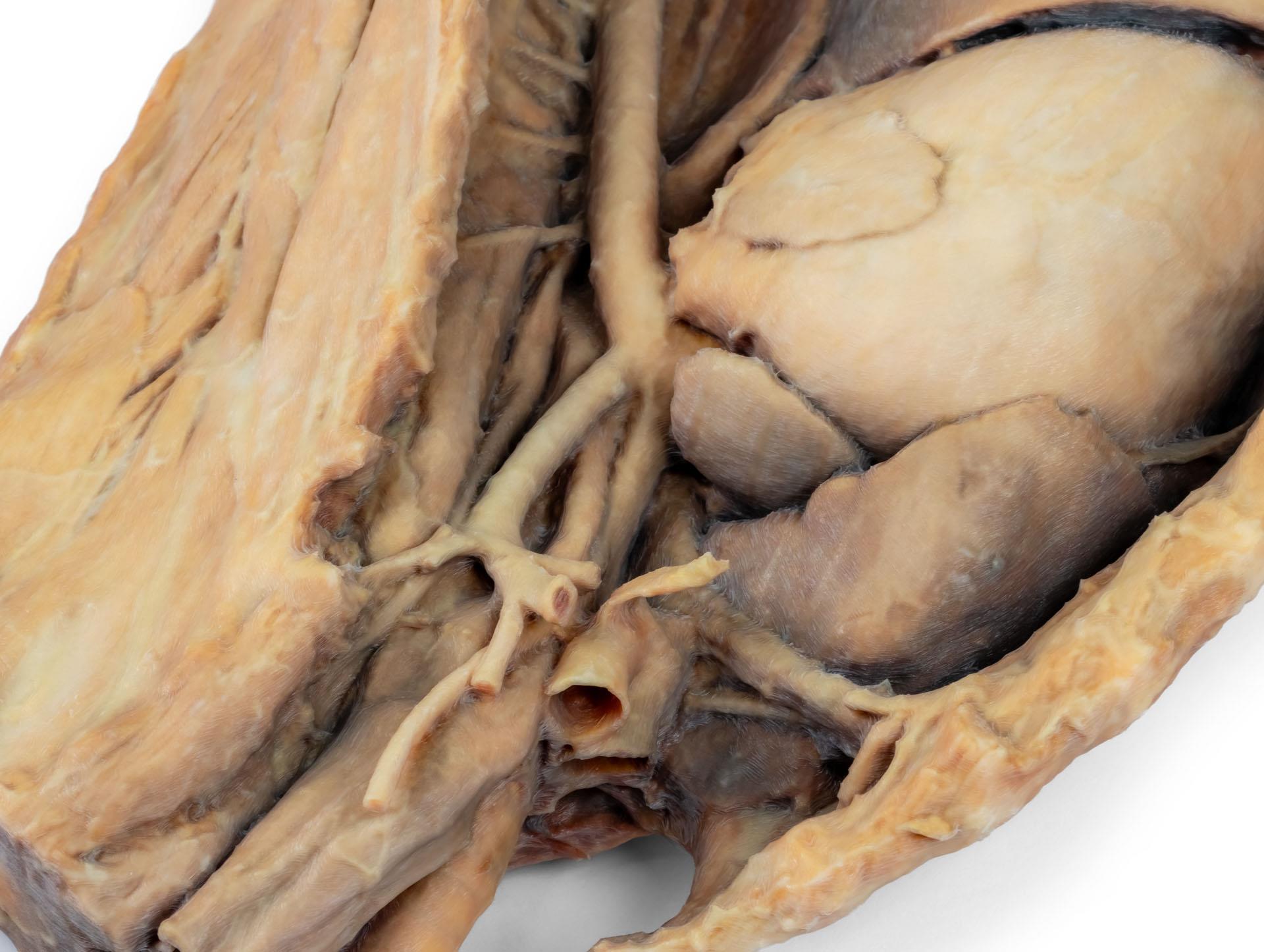
Within the cranial mediastinum, the brachiocephalic trunk and left subclavian artery are visible as major branches arising from the aortic arch, providing key insight into the arterial supply of the thorax and forelimb.
The thoracic oesophagus is visible traversing the mediastinum in a craniocaudal direction, allowing clear identification of its anatomical course through the thoracic cavity.
The preserved diaphragm serves as an important anatomical landmark for retrodiaphragmatic organs such as the liver and stomach. The diaphragmatic crura and their relationship to the aortic hiatus are maintained, along with the initial visceral branches of the abdominal aorta, including:
- Celiac trunk
- Cranial mesenteric artery
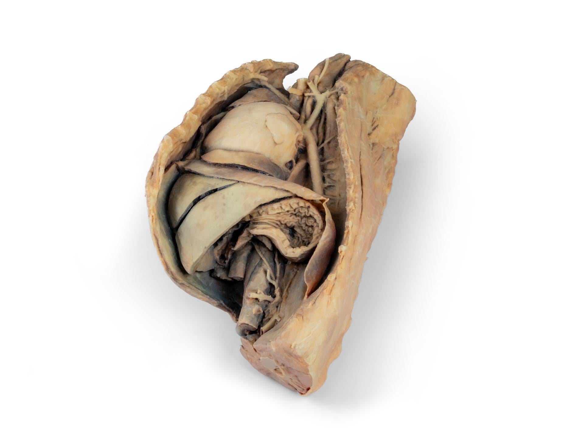
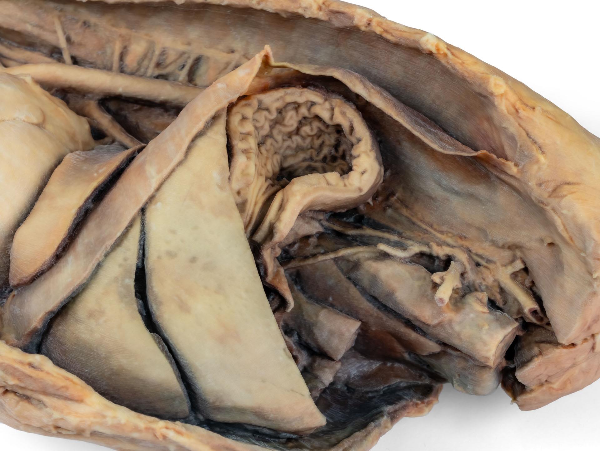
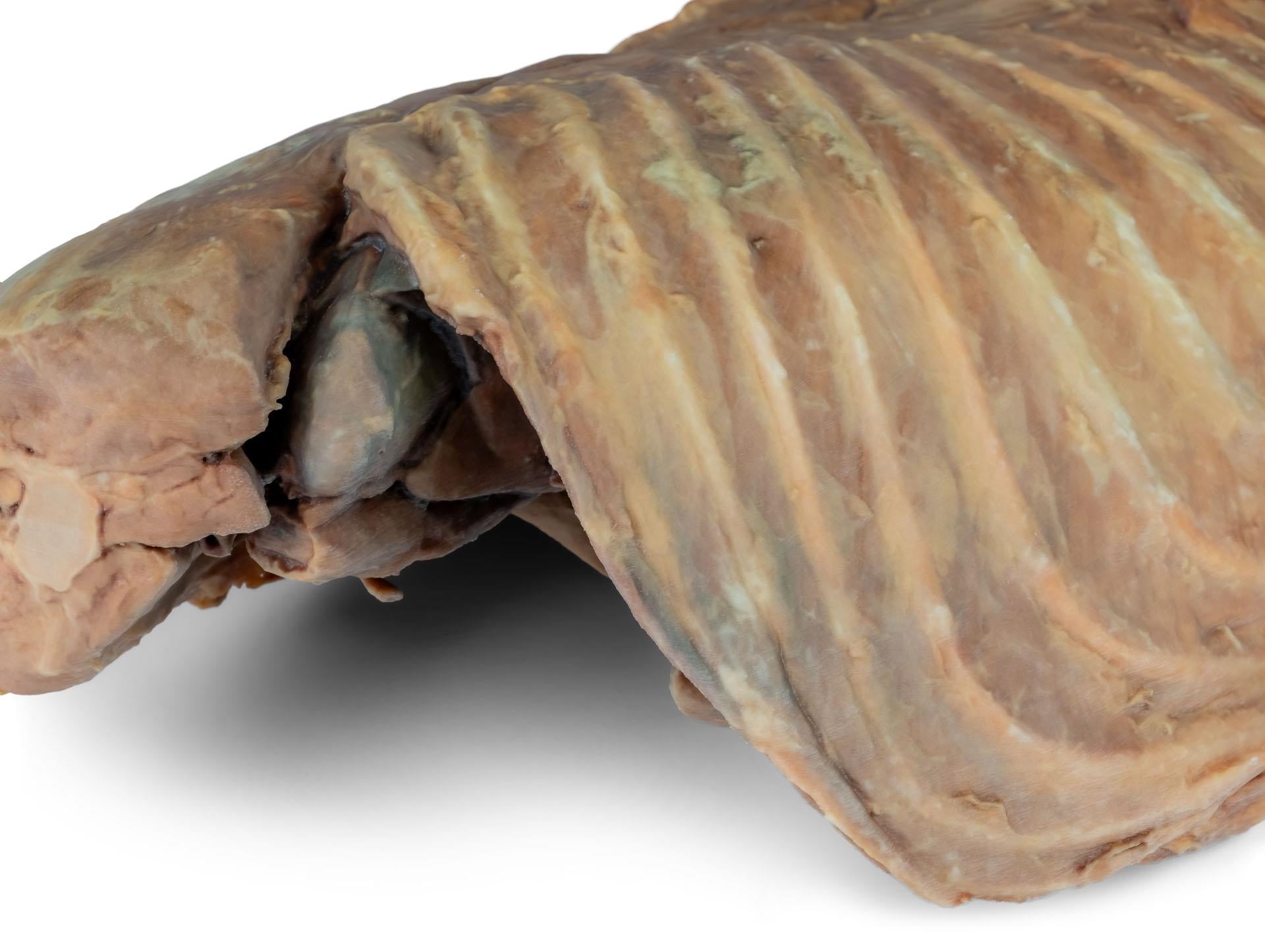
Cardiac structures are also clearly displayed, including the interventricular and coronary grooves. A section of the right atrial wall has been removed to expose the right atrioventricular (tricuspid) valve, including the chordae tendineae and papillary muscles.
Additional internal cardiac features include the septomarginal trabecula (moderator band) within the right ventricle, as well as the outflow tract from the ventricle into the pulmonary trunk. The left atrial wall has been opened to allow clear visualisation of the atrial lumen.
The realistic dissection and comprehensive anatomical detail make this specimen highly suitable for veterinary anatomy laboratories, pathology teaching, and advanced cardiothoracic education.
Key features:
- 3D-printed from a real canine thoracic and cranial abdominal dissection
- Left-sided thoracic cavity approach with preserved anatomical orientation
- Right lung lobes retained as anatomical reference points
- Displays heart, mediastinum, diaphragm, and upper abdominal landmarks
- Visible major vessels, oesophagus, and cardiac internal structures
- Ideal for advanced veterinary anatomy and surgical training
Ideal for veterinary schools and universities for anatomy and pathology instruction as well as clinical and surgical training.