


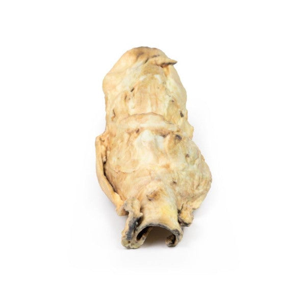


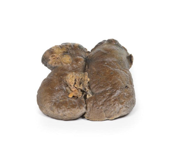
Carcinoma of Larynx
The specimen includes the tongue, pharynx, larynx, oesophagus, and trachea, presented in the coronal plane. The opened oesophagus and trachea reveal a fungating carcinoma, extending into both pyriform fossae. The tumour, originating from the larynx, affects both vocal cords, the left aryepiglottic fold, and both pyriform fossae, displaying an irregular surface with areas of shaggy necrosis.
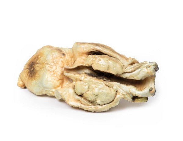
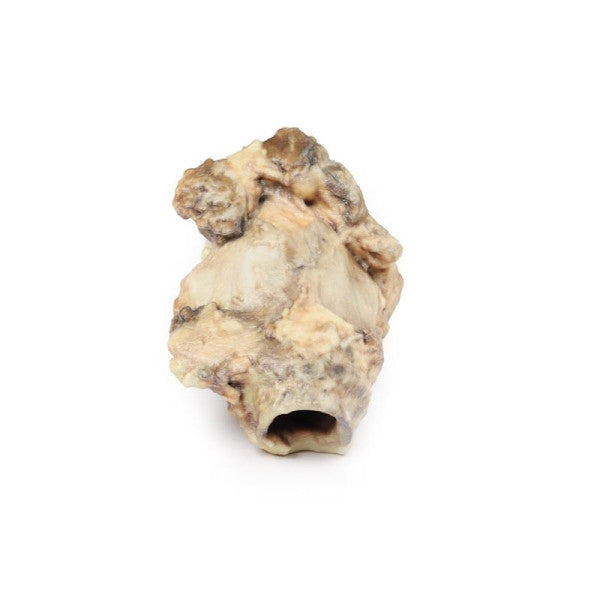
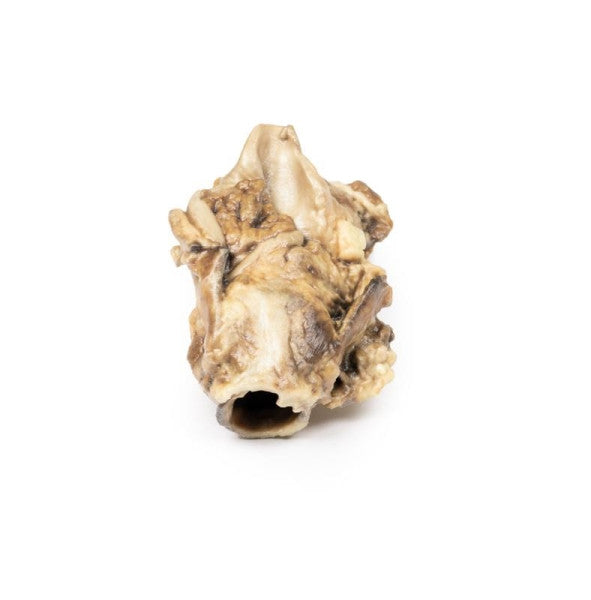
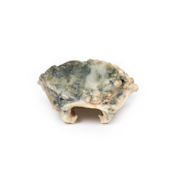
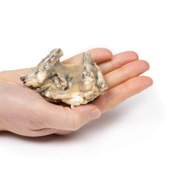
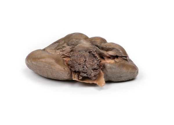
Carcinoma of Pyriform Fossa
The amputated larynx, observed from the rear, exhibits an irregular and fungating tumour originating in the left pyriform fossa. The laryngeal tissues display distortion and oedema. Upon histological examination, the tumour was identified as squamous cell carcinoma.
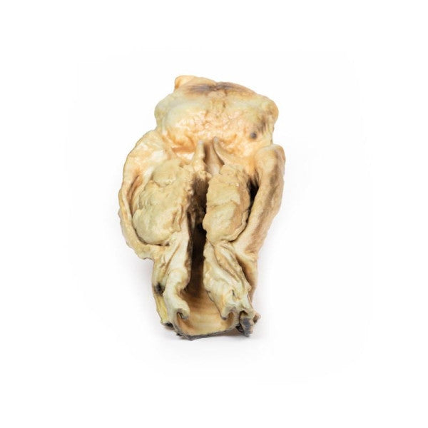
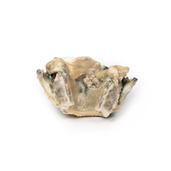
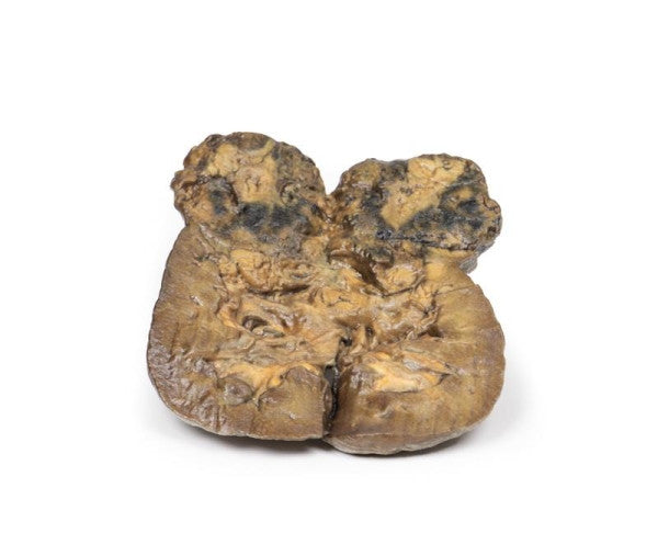
Carcinoma of Larynx (Posterior Aspect)
This specimen represents a patient's laryngectomy, with the larynx cut open and observed from the posterior view. A noticeable irregular ulcerating tumour causes significant distortion to the right vocal cord, accompanied by mucosal congestion. Histologically, the identified pathology is a well-differentiated squamous cell carcinoma.
Carcinoma of Breast
This specimen is the left breast presented to showcase its cut surface. A substantial oval tumour is situated just beneath and adherent to the skin, extending into the underlying muscle. The tumour lacks encapsulation and exhibits a diverse cut surface with regions of necrosis, haemorrhage, and cyst formation. The identified pathology is a breast adenocarcinoma that has also affected the nearby lymph nodes.
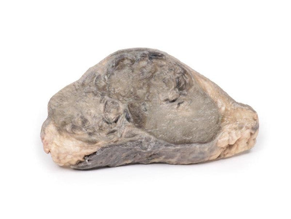
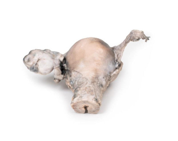
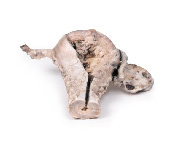
Endometrial Carcinoma
The specimen includes the uterus, fallopian tubes, and ovaries. The anterior aspect reveals an abnormal endometrial lining, particularly on the right side, with a brown polypoid tumour invading the myometrium and extending into the cervical canal. Histologically, this was identified as a well-differentiated adenocarcinoma of the endometrium. The left ovary, sectioned coronally, exhibits enlargement and several sizable follicular cysts/cavities.
Hepatocellular Carcinoma
This liver specimen from a postmortem examination exhibits a multinodular appearance, indicative of macronodular cirrhosis. Multiple nodules are dispersed with narrow fibrous bands. Additionally, two large round tumours display a variegated cut surface marked by focal necrosis, haemorrhage, and bile staining. This illustrates hepatocellular carcinoma development within a cirrhotic liver.
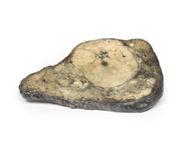
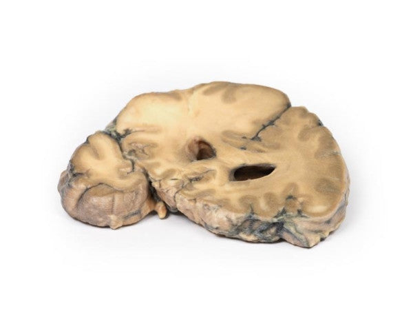
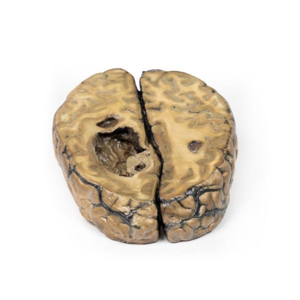
Metastatic Adenocarcinoma in the Brain
This brain specimen, sectioned coronally, reveals a distinct pink-grey tumour located in the right frontal lobe, affecting both grey and white matter. The tumour is circumscribed and exhibits variegation. Its presence causes compression of the right lateral ventricle and a noticeable shift in midline structures.
Metastatic Carcinoma
The left lung, sliced longitudinally exhibits numerous dispersed pale tumour nodules of different sizes. Some nodules near the hilum merge and the hilar lymph nodes contain pale tumour tissue. Small nodules are visible beneath the thickened pleura. Histologically, these are metastatic deposits of adenocarcinoma. Post-mortem findings revealed ovarian adenocarcinoma with metastases in the lungs, heart, liver, and pericardium.
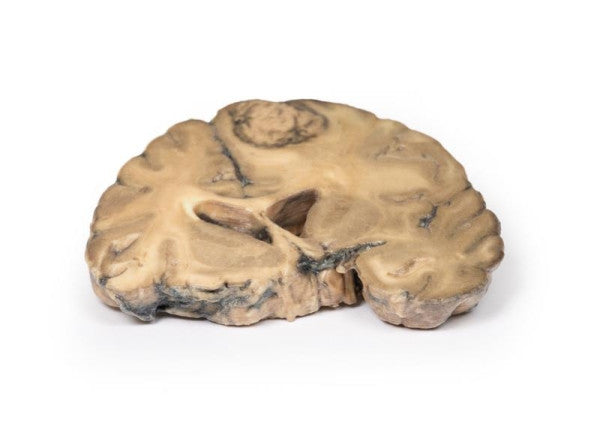
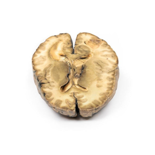
Metastatic Carcinoma in the Brain
The cerebrum specimen shows an enlarged right hemisphere with three cystic tumours, the largest being 5 cm in the right parietal region. These tumours, exhibiting shaggy greyish tissue, had metastatic carcinoma, with liver and bone metastases from primary breast carcinoma.
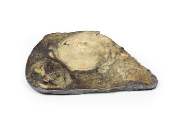
Papillary Transitional Cell Carcinoma of the Renal Pelvis
This kidney, post-nephrectomy, retains its foetal lobulation. It exhibits a friable papillary tumour projecting into the renal pelvis, causing visible dilatation. Histological analysis confirmed it as papillary transitional cell carcinoma originating in the renal pelvis.
Renal Cell Carcinoma
This kidney specimen, dissected coronally, reveals an irregular lower pole tumour causing parenchymal compression. The cut surface shows variegation, with intrarenal metastases as small pale-yellow nodules. The renal pelvis exhibits slight dilatation and papillary blunting, suggesting mild hydronephrosis. The capsular surface is nodular, with scars and small cysts. Histologically, it is diagnosed as renal cell carcinoma.