


Our Chicken Anatomy Poster is a high-quality educational wall chart featuring accurate, full-colour illustrations of the chicken’s anatomy. Designed by our specialist medical and veterinary illustrators, this chart is the perfect learning resource for veterinary students, poultry farmers, agricultural colleges, and school science classrooms.
The poster provides a clear overview of chicken biology with labelled diagrams covering:
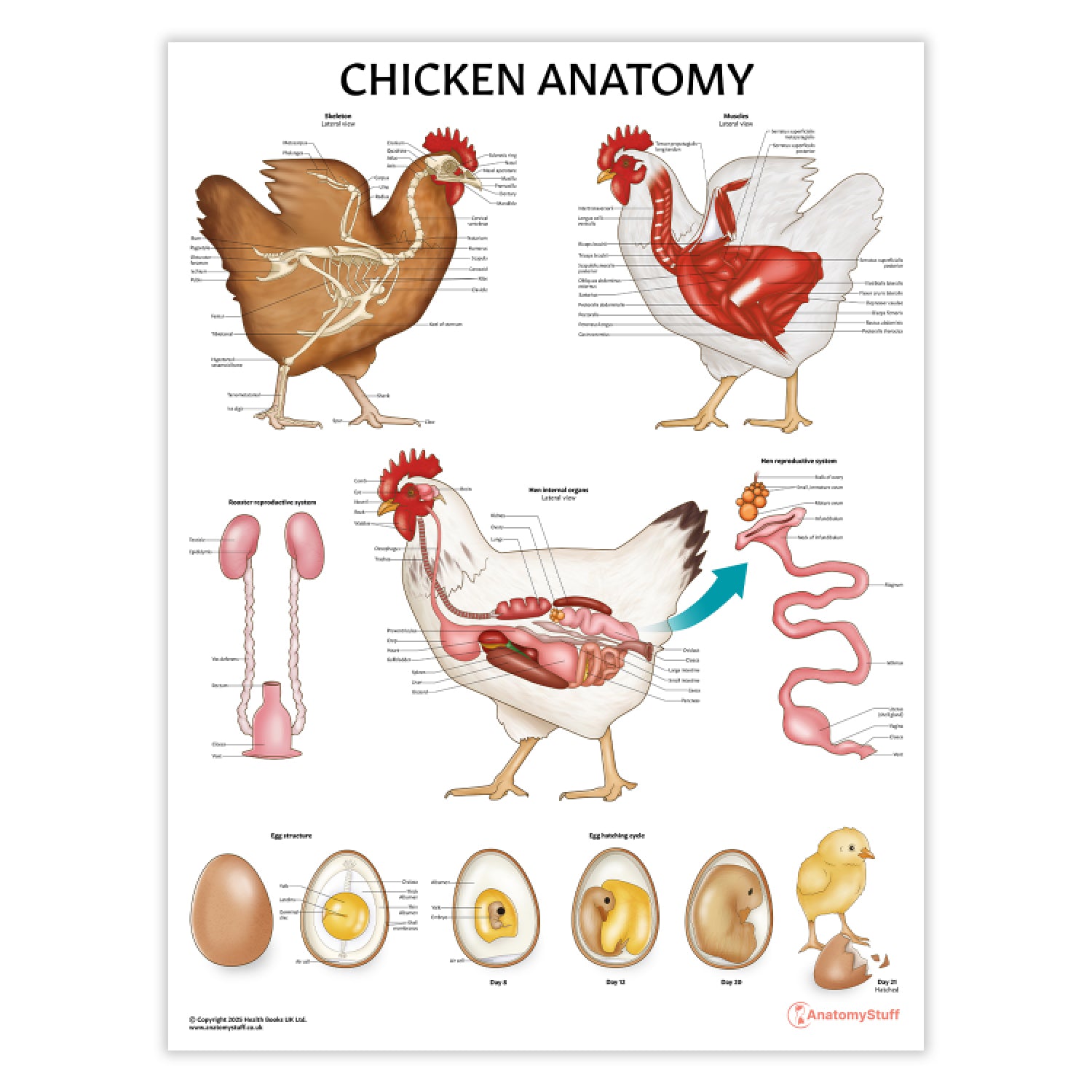
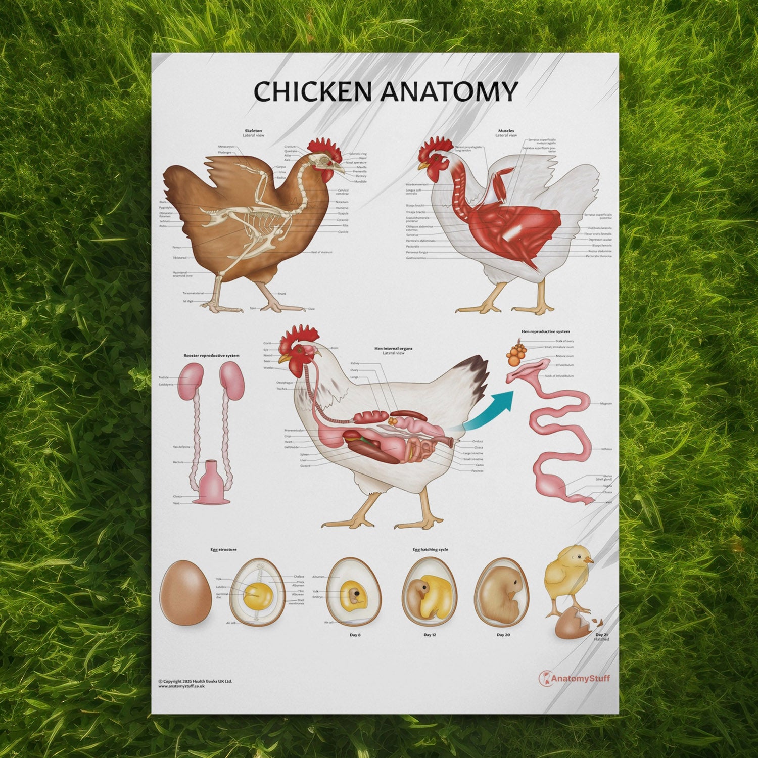
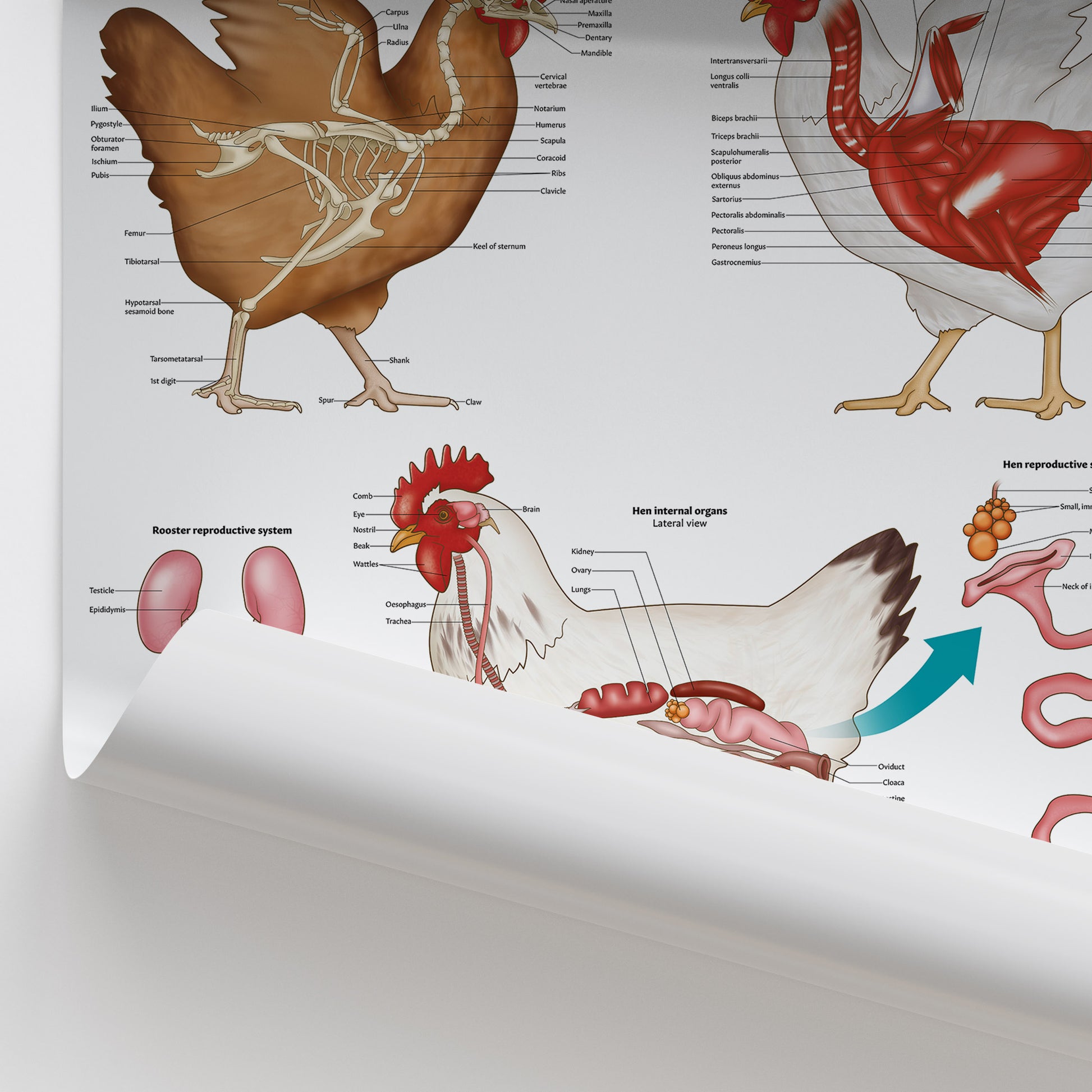
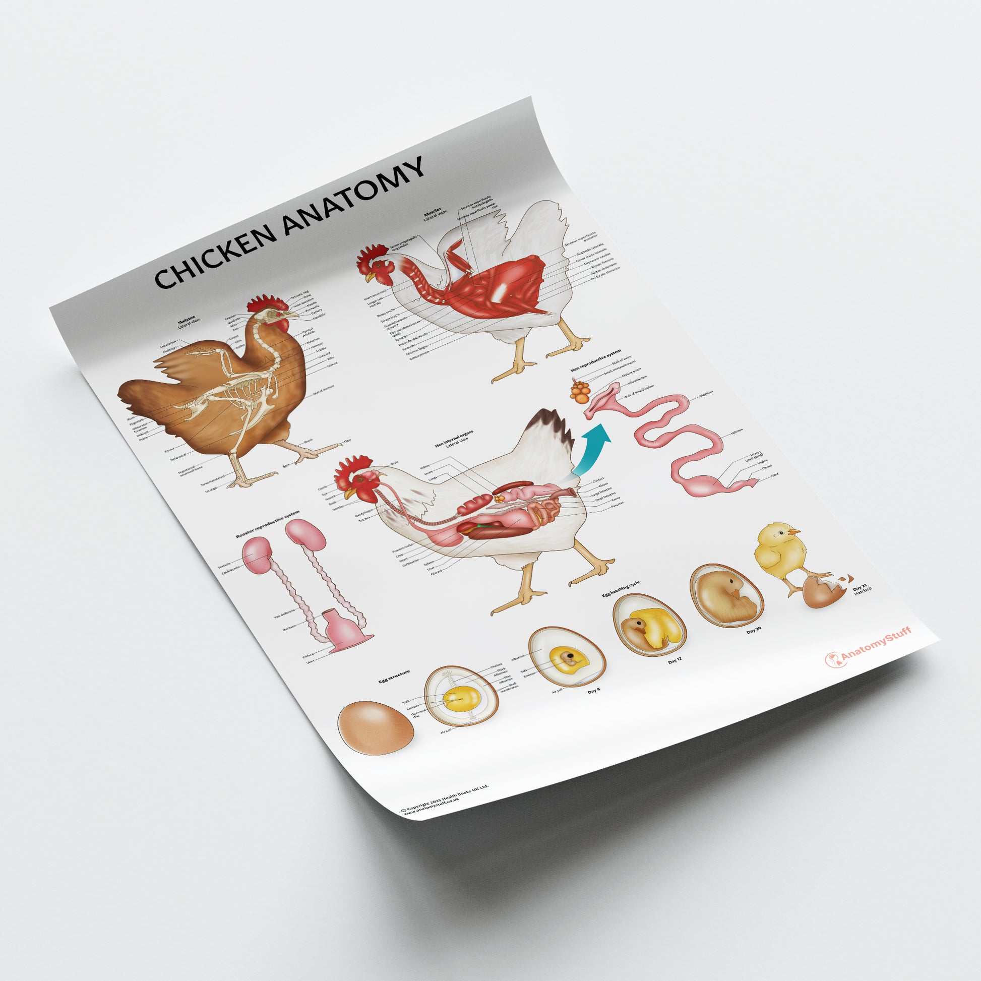
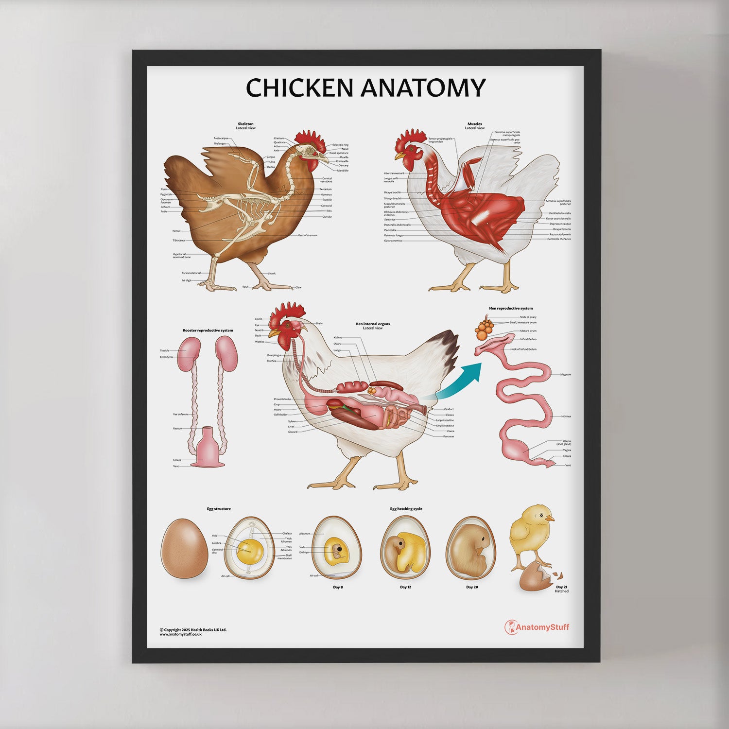
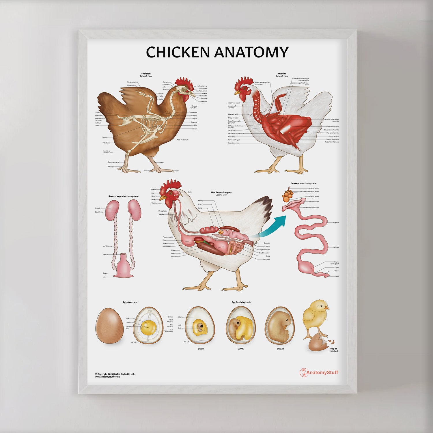
- Skeletal anatomy – lateral view showing major bones such as the sternum, femur, and skull
- Muscular anatomy – detailed view of key muscles including pectorals and gastrocnemius
- Internal organs – digestive, respiratory, and circulatory systems illustrated in situ
- Hen reproductive system – ovary to cloaca, with egg development pathway shown
- Rooster reproductive system – labelled diagram of testes and vas deferens
- Egg structure and hatching cycle – from egg formation to chick development and hatching
This chicken anatomy wall chart is ideal for:
- Veterinary and agricultural training centres
- Poultry farmers seeking a quick reference guide
- Secondary schools and higher education classrooms
- Farm education centres and visitor displays
Our collection offers a range of display options to meet your needs:
- Classic Semi-Glossy Prints: Available in 45 x 60 cm, 60 x 80 cm, and 70 x 100 cm sizes. The semi-glossy finish enhances colours with a subtle shine, adding vibrancy to any setting.
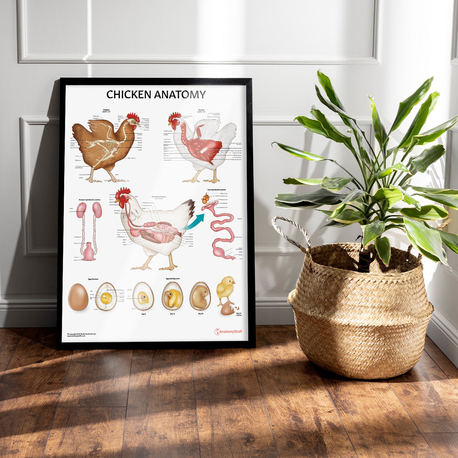
- Framed Prints: Offered in black or white frames and sizes 45 x 60 cm, 60 x 80 cm, and 70 x 100 cm. Features 170 gsm matte paper with a smooth, non-reflective finish. Ready-to-hang with a durable pine wood frame.
