


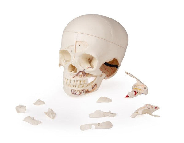
This Child's Demonstration Skull Model has been cast from a real human child skull specimen, to ensure it shows all of the anatomical structure in detail. This skull has been complexly cut and joined back together with metal and magnetic connections. This skull is modelled on an estimated age of a 3-year-old child.
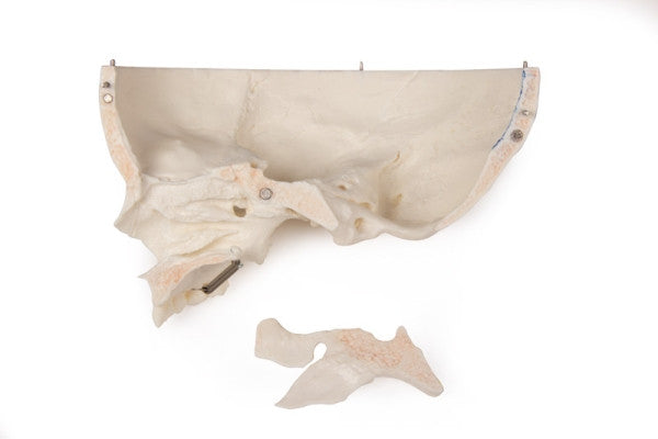
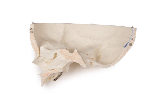
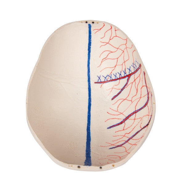
The top of the skull can be opened and is also removable. Bony impressions of the sinus sagittalis, the sinus transversus, the sinus sigmoideus and the meninges are painted to highlight the internal details.
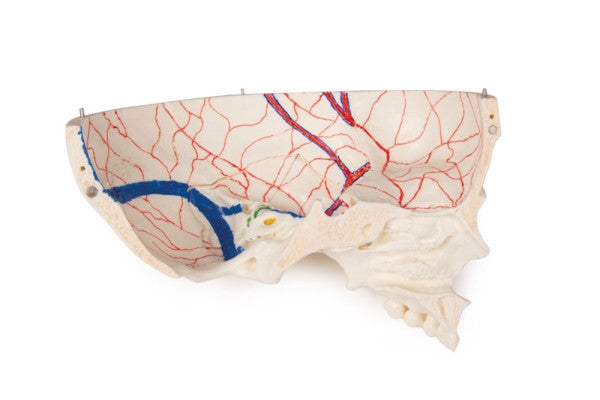
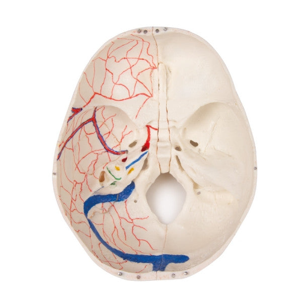
The base of the skull has been cut sagittally, the cut runs through a sieve plate on one side. Another cut has been made with the same plane through to the other sieve plate of the ethmoid bone, leaving the Christa galli, the perpendicular plate and the entire nasal septum in its original state. The anterior, middle and posterior fossa structures are easily accessible. Students and medical professionals can see the nasal cavity, the turbinate, the septum and the bony nasopharynx directly. The nasal septum can be removed from the surrounding bone structures. The removable temporal bone shows a complete external auditory canal.
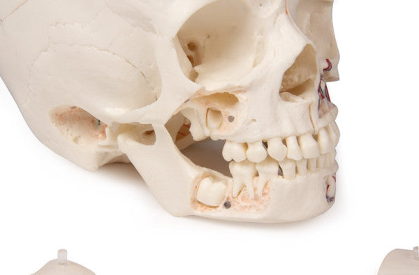
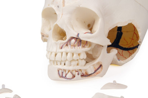
The upper jaw and the lower jaw show the structures of the teeth, the roots, the bony edge of the alveolar process, as well as dental nerves and vessels. The maxillary sinus can be opened by removing a flap. The teeth, including the permanent teeth still embedded in the bone, can be made accessible by removing a bone flap.
Developed for students of anatomy, medicine, surgery, ENT medicine, ophthalmology and dentistry.