


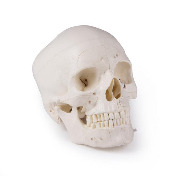
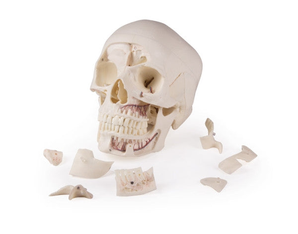
This life-like Demonstration Skull Model has been created by casting a real human specimen to ensure the highest of detail and accurate anatomical structures.
This Erler Zimmer 14 Piece Demonstration Skull has been intricately sectioned and then reassembled with metal and magnet connections, making it the perfect demonstration aid.
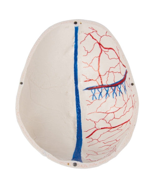
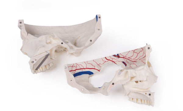
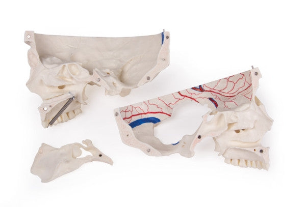
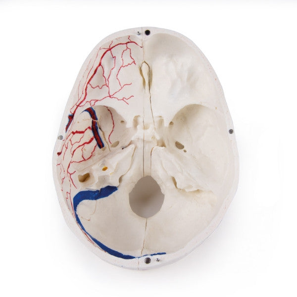
The temporal bone and its sutures are intact, with the calvarium being sectioned horizontally. The bony impressions of the superior sagittal sinus, the transverse sinus, the sigmoid sinus as well as the meningeal vessels have been painted to highlight the detail.
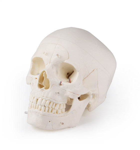
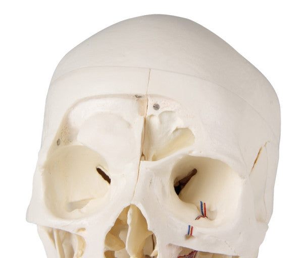
This skull has been sectioned sagittally in the way that it passes through the cribriform plate on one side, and another section, in the same way the plane passes through the other cribriform plate of the ethmoid. This leaves the crista galli perpendicular plate of the ethmoid intact as well as the whole nasal septum.
Anterior, middle and posterior cranial fossae structures are easily accessible on this model. Students and professionals will be able to visualise the nasal cavity, the concha, the nasal septum, the bony pharyngeal and also nasopharyngeal spaces.
On one side the frontal sinuses have been chiselled out for full access to the sinus, on the other they have been dissected to show the sinus as a whole. The relation of the sinus to the nasal cavity is clearly shown. In addition to this the nasal septum is separable from the surrounding bones.
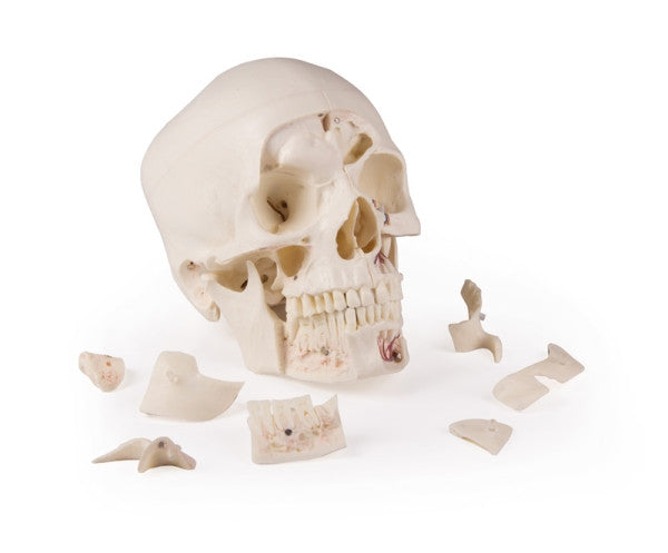
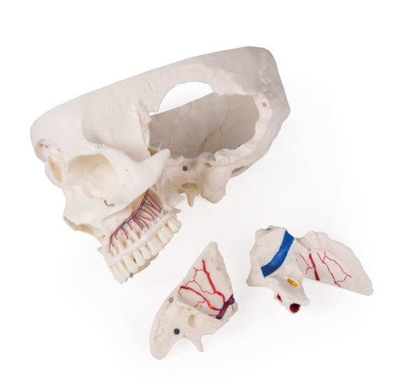
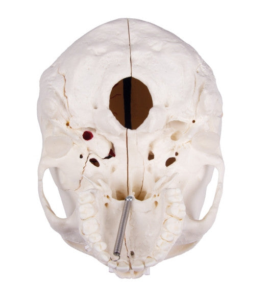
On one side of the skull model the temporal bone has been left in its original position. The other temporal bone is removable from the skull. A section of the mastoid and squama can be removed, along with the tympanic antrum, allowing the internal ear to be viewed in full.
All three semicircular canals are visible, with the facial nerve coursing backwards and downwards, finally appearing through the stylo-mastoid foramen. The removable temporal bone has the external auditory meatus intact. The vertical section through the squama mastoid carrying inwards along the petro-squamosal junction has been created and when apart, students can see the position of tympanic membrane. The carotid canal has been opened up, as well as the cochlea, showing that the internal canal, and the course of the facial nerve has been depicted. Oval windows, semi-circular canals, and aditus of the tympanic antrum are visible.
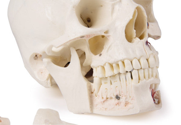
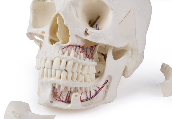
The maxilla and mandible expose the structures of dentition. The roots, bony margin of the alveolar process, dental vessels and nerves are all visible. The maxillary sinus can be opened by removing the bone flap. Teeth of the right mandible are sectioned to show the inner tooth structure.
This Demonstration Skull Model is ideal for students studying anatomy, medicine, surgery, otolaryngology, ophthalmology and dentistry.