


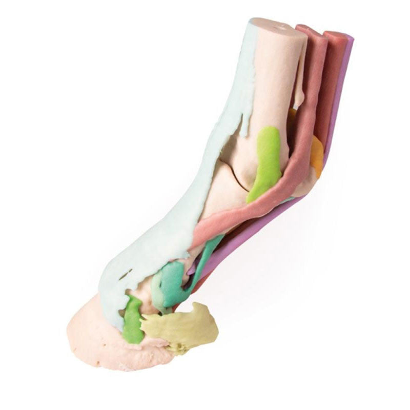

This Equine Hoof 3D Printed Model - Model 1 has been carefully created based on CT and MR co-registered data.
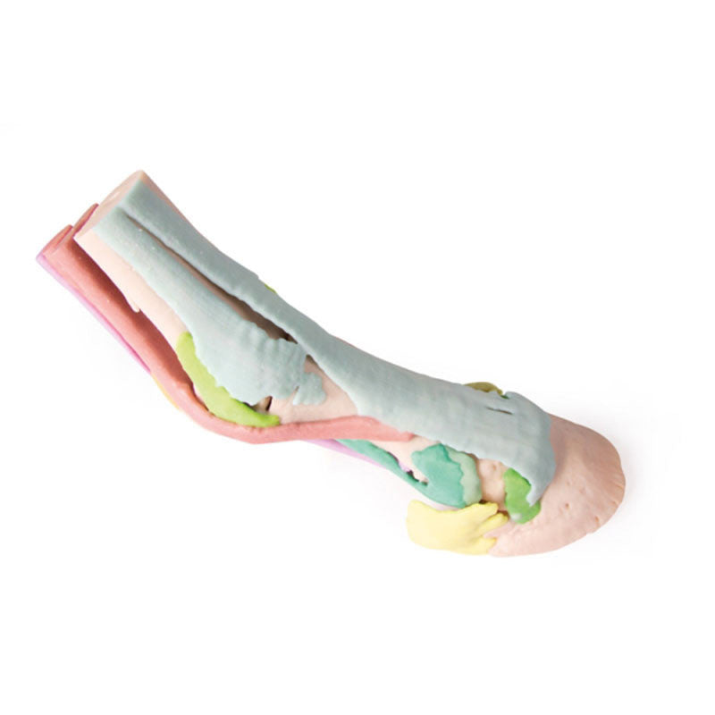
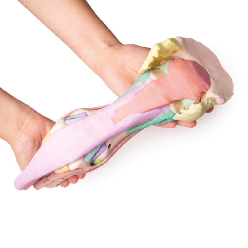
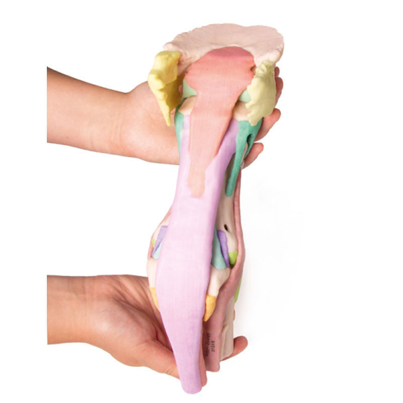
Anatomically accurate and life-sized, this full-colour 3D printed model makes for the perfect equine training tool. This horse hoof model shows the full anatomy of a horse's foot and is made up of 25 anatomical structures.
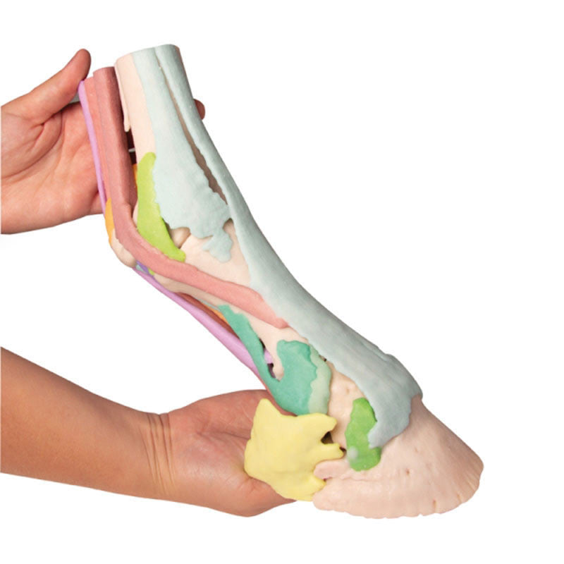
Each anatomical component is uniquely coloured, helping vet and equine students to easily recognise them.
This Equine Hoof 3D Printed Model is part of a series from Erler Zimmer which consists of four models. These range from models showing anatomical structures to models showing deeper structures.
Model 1 includes the following structures:
Third metacarpal bone; Proximal sesamoid bones; Proximal phalanx; Middle phalanx; Distal phalanx; Distal sesamoid (navicular) bone; Ungular cartilages; Collateral ligaments of the metacarpophalangeal joint; Collateral ligaments of the proximal interphalangeal joint and the abaxial palmar ligaments of the foot; Collateral ligaments of the distal interphalangeal joint; Collateral ligament of the distal sesamoid (navicular) bone; Distal sesamoidean impar ligament; Proximal scutum and intersesamoidean ligament; Cruciate sesamoidean ligament; Short sesamoidean ligaments; Oblique sesamoidean ligaments; Straight sesamoidean ligament; Axial palmar ligaments of the foot; Suspensory ligament and extensor branches; Deep digital flexor tendon; Superficial digital flexor tendon; Common digital extensor tendon.
Hoof capsules can be purchased separately, helping to anchor the distal limb in the stance position.