


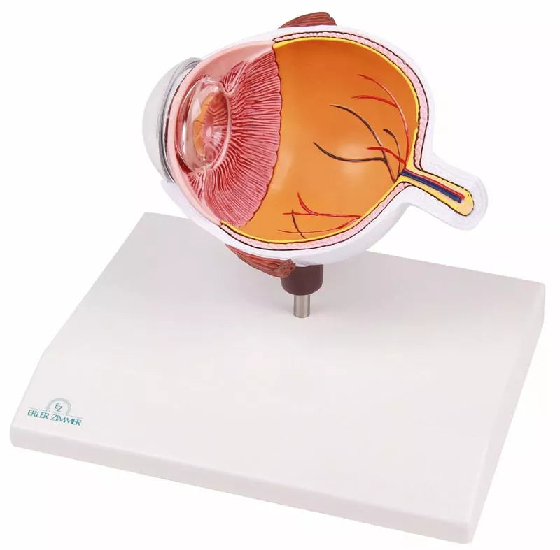
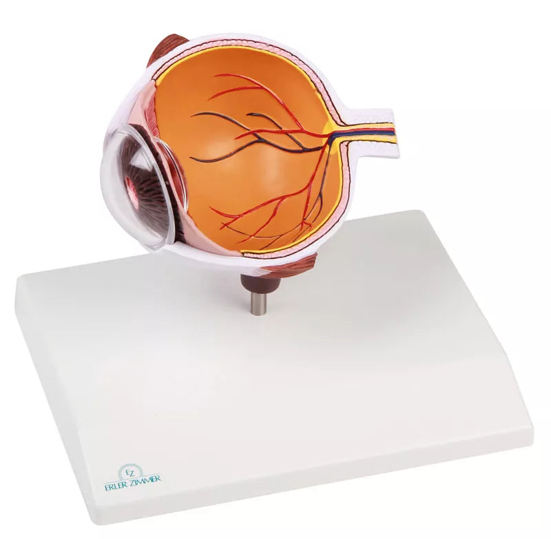
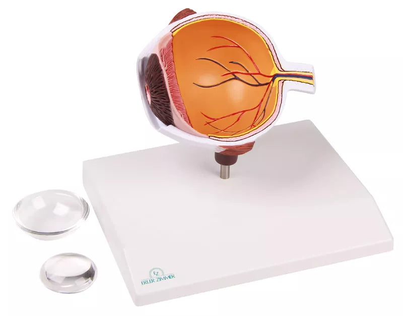
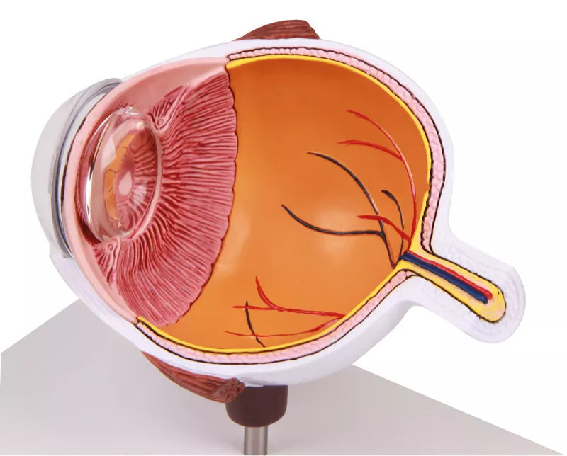
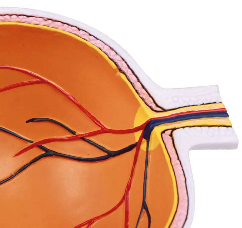
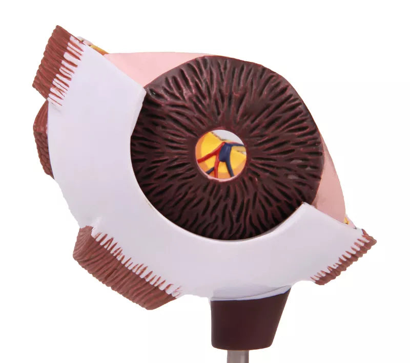
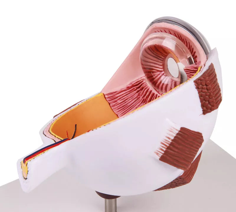
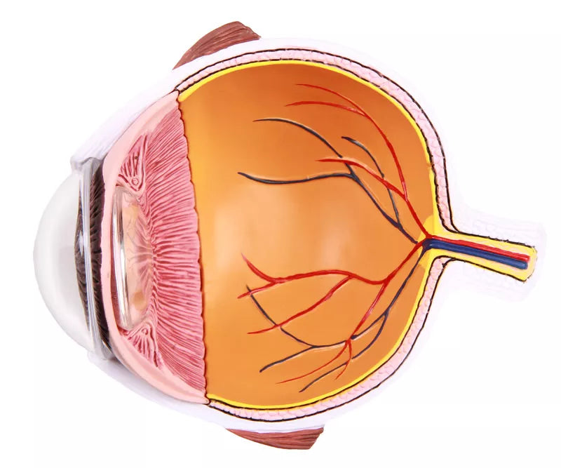
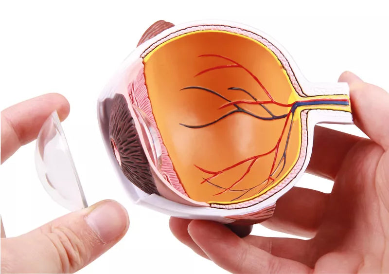
This Human Eye Half Model presents a detailed cross-section through the human eyeball, enlarged for optimal study of ocular anatomy. The model accurately illustrates the intricate structures of the eye, making it an excellent tool for both education and patient explanation.
The following anatomical features are clearly displayed: choroid, retina, macula, optic disc, optic nerve, central retinal artery and vein, retinal blood vessels, superior and inferior rectus muscles, ora serrata, lens, iris, cornea, and sclera. Each structure is carefully coloured and detailed to enhance visual understanding of the eye’s complex anatomy.
Augmented Anatomy App Included
Enhance your learning experience with the innovative Augmented Anatomy App, designed to work seamlessly with this high-quality model. Simply point your device at the model, and the app automatically recognizes it, displaying anatomical labels and terminology in real-time augmented reality.
App Features
- High-quality augmented reality learning tool
- Free to use, with no registration required
- Anatomical nomenclature available anytime and anywhere
- Additional online resources included in the learning lexicon
- Compatible with all major smartphones and tablets
This model is ideal for anatomical study and display in GP clinics, ophthalmology practices, hospitals and universities.