


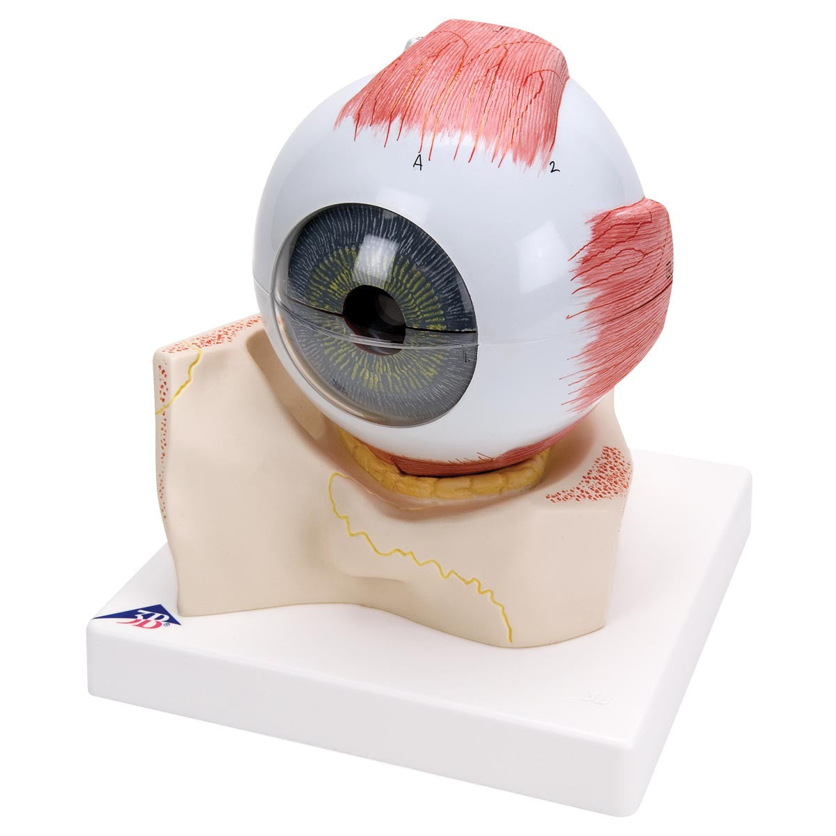
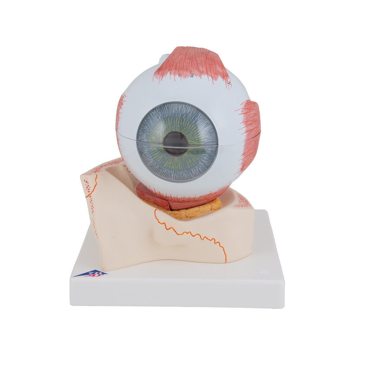
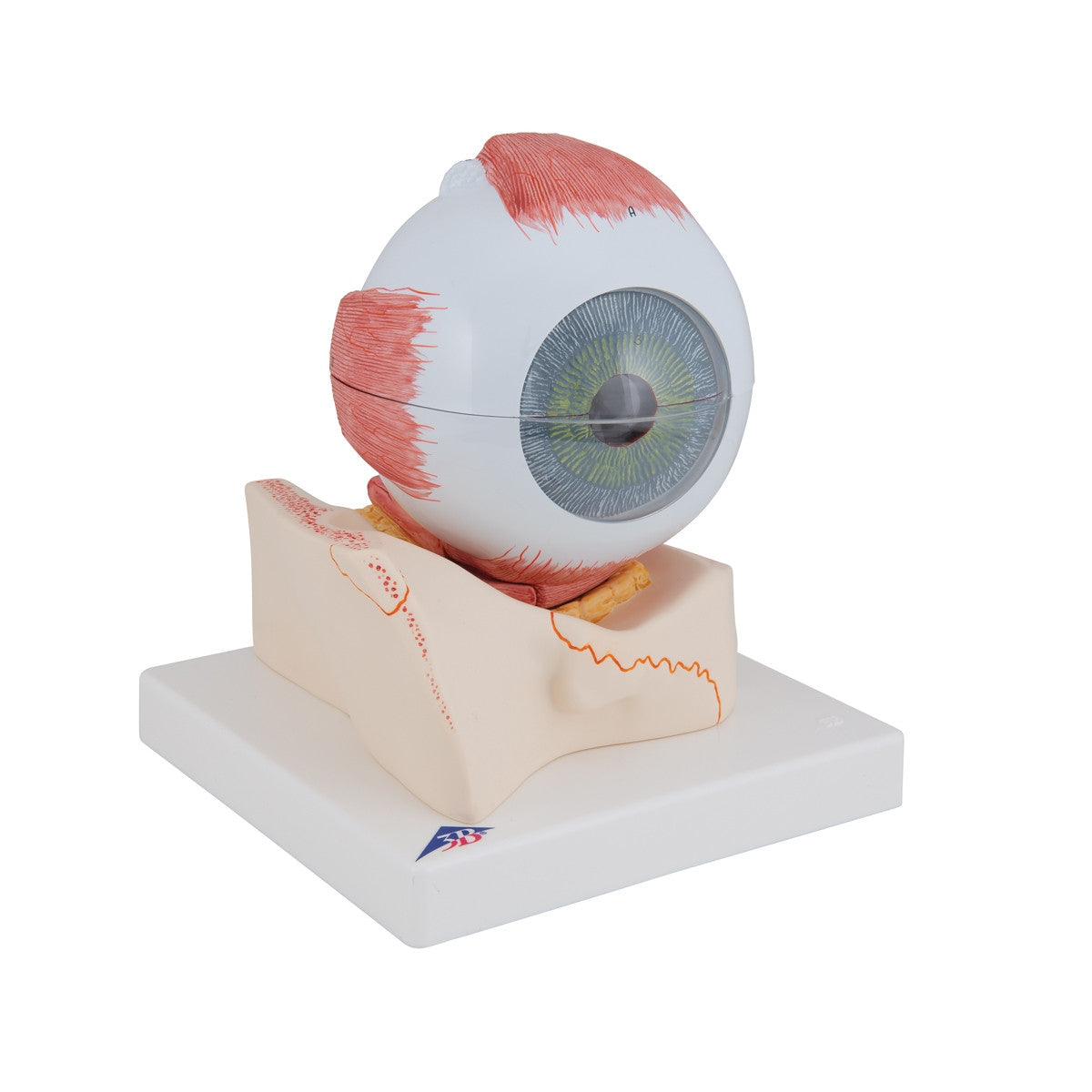
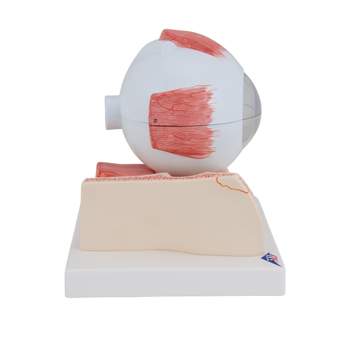
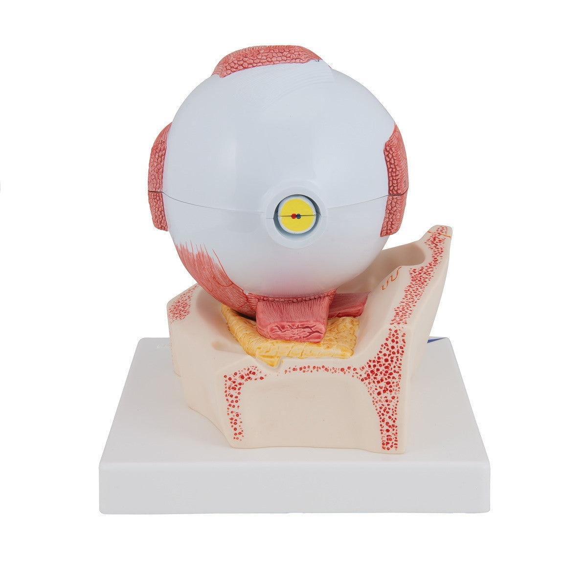
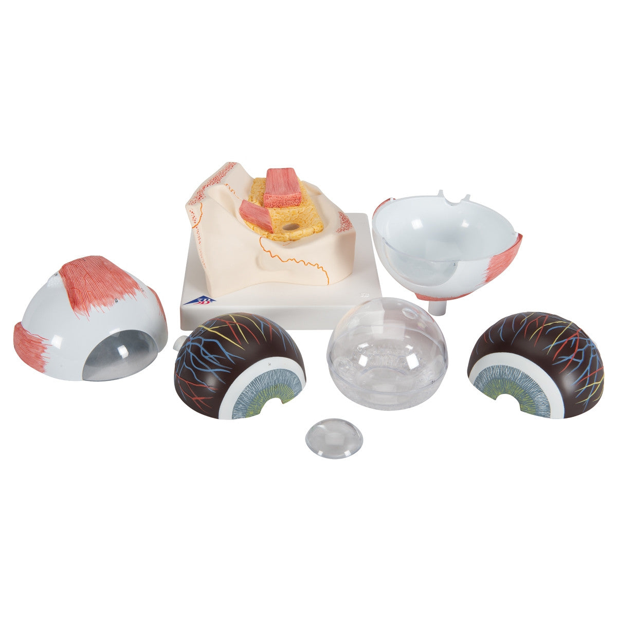
This large human eye model by 3B Scientific can be dismantled into seven parts for closer study and is five times life-size. The removable eye structures include:
- vitreous humour
- the lens
- both halves of chorid with iris and retina
- the upper half of the sclera with cornea
- eye muscle attachments.
The F11 / 1000256 model eye is set on the base of bony orbit. This enlarged eye model would be ideal as a teaching aid in a student classroom or lecture hall.
The following anatomical features appear on this eye model and are listed in English and Latin on the keycard provided:
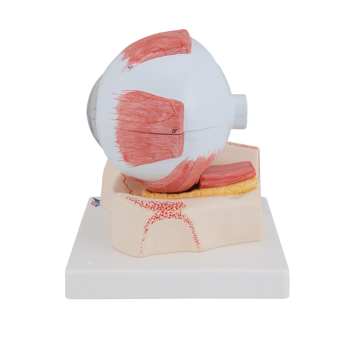
Extra-ocular muscles
I Superior rectus
II Inferior rectus
I II Medial rectus
IV Lateral rectus
V Tendon of superior oblique
VI Inferior oblique
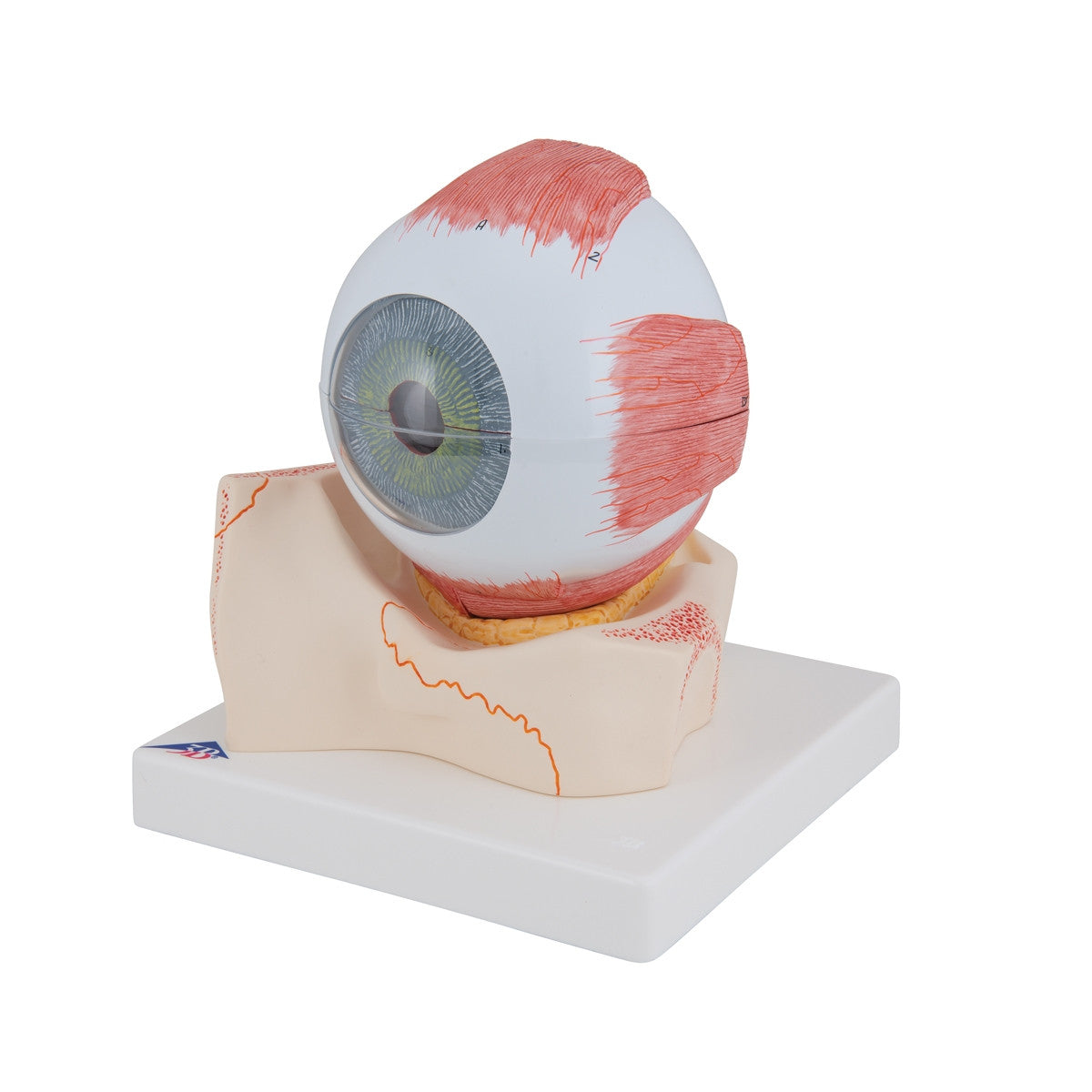
A Fibrous layer of eyeball
1 Cornea
2 Sclera
B Vascular layer of eyeball
3 Iris with pupil
4 Ciliary muscle
5 Corona ciliaris
6 Choroid
7 Ciliary nerves
8 Vorticose vein
8a Ciliary arteries
C Inner layer of eyeball
9 Retina
10 Ciliary part of retina
11 Retinal arterioles
12 Retinal venules
13 Macula and fovea centralis
14 Optic disc
15 Lens
16 Vitreous body
Download Eye Model (5 times life size, 7 part) F11 / 1000256 product manual here.
This model comes with 3B Scientific 3B Smart Anatomy app included for FREE. This features access to an anatomy course, including 3 digital anatomy lectures, 117 different virtual anatomy models and 39 anatomy quizzes. It also offers a FREE warranty upgrade from 3 to 5 years with every product registration. To unlock these benefits, scan the label located on your 3B Scientific anatomy model and register online.