


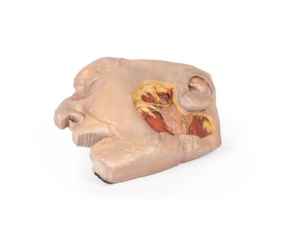
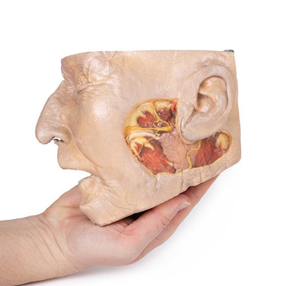
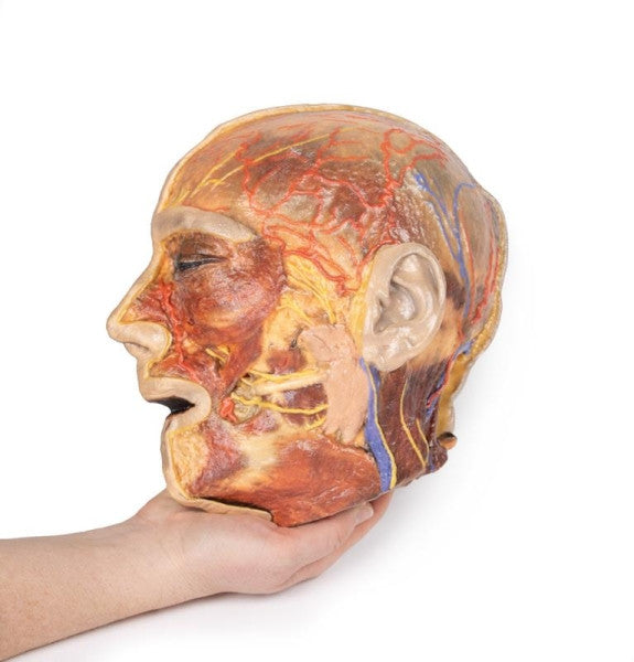
Parotid Gland and Facial Nerve Dissection
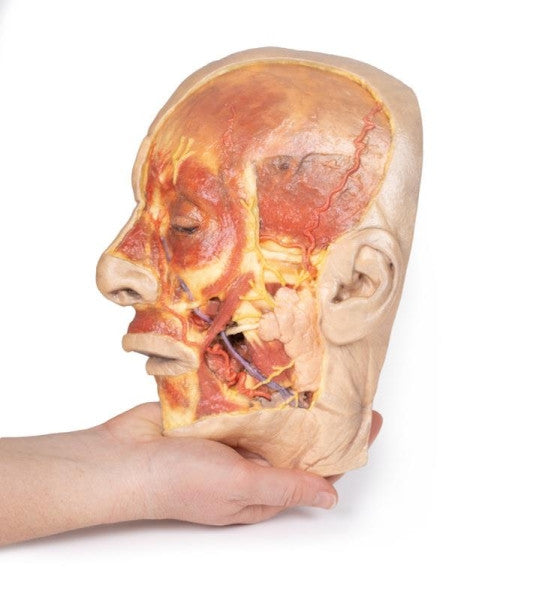
This 3D model showcases parotid gland anatomy in a lateral face dissection, vital for procedures like Mohs surgery. The window spans from the anterior external ear to the mandibular angle, exposing key structures such as the superficial temporal artery, facial nerve branches, parotid duct, and the ascending branch of the great auricular nerve.
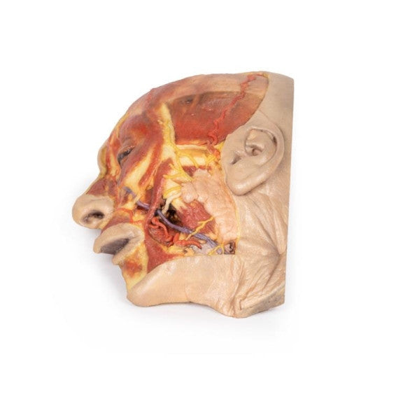
Superficial Face
This 3D model offers a superficial dissection of the left face anterior to the ear, emphasising neurovascular structures and facial muscles. The dissection window reveals the parotid gland, facial nerve branches, facial artery, and vein. Tracking these structures highlights muscles like the masseter, depressor anguli oris, zygomaticus, and orbicularis oculi, providing a comprehensive view of facial anatomy.
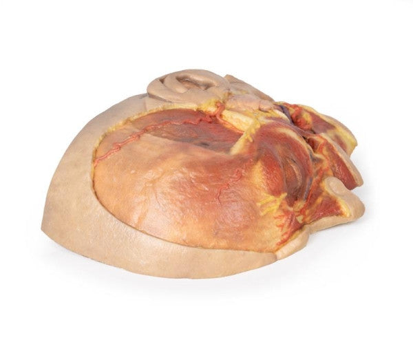
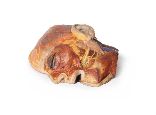
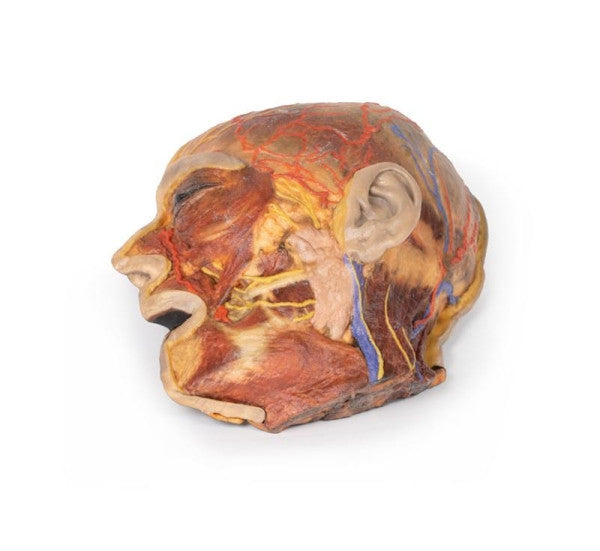
Superficial Facial Nerves & Parotid Gland
This 3D model offers an expanded dissection of the facial and occipital regions, with the dissection being extended below the ear. The model showcases neurovascular details, such as the supraorbital and supratrochlear nerves and arteries. The retromandibular vein, great auricular nerve, and occipital artery are visible, providing a comprehensive view of the head's superficial anatomy.
Face 3D Printed Anatomy Model - MP1112-108-109
Dimensions: 36 cm x 26 cm x 15 cm
Weight: 2.5 kg
Manufacturer: Erler Zimmer
Made in Germany