


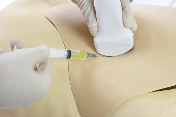
Experience a real-life representation of an ultrasound-guided femoral arteriovenous puncture and abdominocentesis procedure, with this Femoral Arteriovenous Punction and Abdominocentesis Ultrasound Training Model. This is a great asset for medical education, practical skills training, as well as radio-technology training, fit for the use of a wide variety of medical professionals.
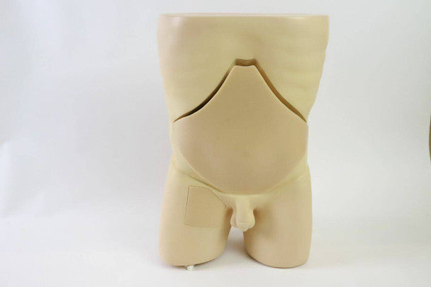
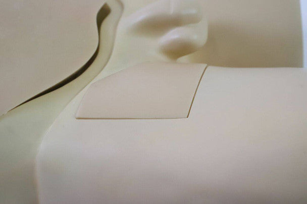
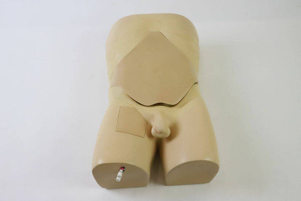
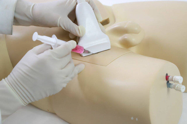
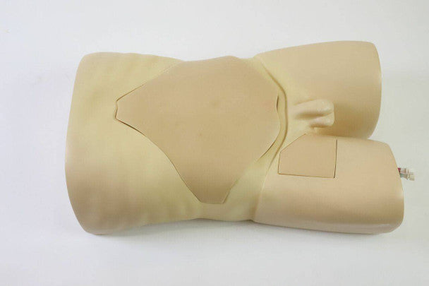
Distinct anatomical landmarks are easily identifiable such as the: abdomen, anterior superior iliac spine, navel, symphysis pubis, groin, femoral artery and femoral veins. High-quality materials have been used for a true-to-life skin touch, feel and puncture, where a resistance and “pop” can be felt with each needle insertion.
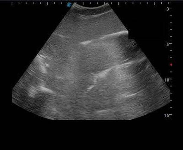
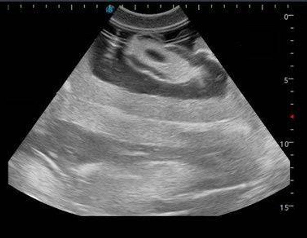
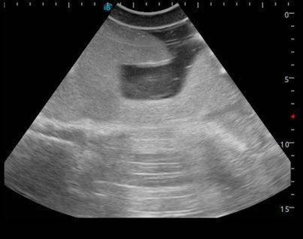
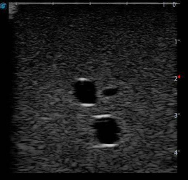
The model emulates real clinical ultrasound images of anatomical structures like the intestines, liver, arteries and veins, and is compatible with various actual ultrasound machine models. Students are thus able to learn how to recognise landmarks by ultrasound and image interpretation. Some of the clinical skills which can be gained are:
- The ability to palpate anatomical landmarks
- The use of ultrasounds and how to interpret them
- Abdominal percussion to determine size and density of structures and organs
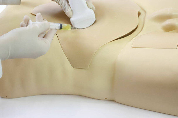
- Abdominocentesis
- Femoral arteriovenous puncture
The Femoral Arteriovenous Puncture and Abdominocentesis Ultrasound Training Model is a versatile teaching tool for the training of various clinical techniques. The resistance of the model to needle tips and the correct anatomical landmarks provide an overall excellent hands-on experience.
Contents Included
1 x Femoral arteriovenous puncture and abdominocentesis ultrasound training model
1 x 150ml Red contrast solution
1 x 150ml Blue contrast solution
3 x 500ml Yellow contrast solution
1 x 200ml Syringe
1 x Water inlet connector
1 x Operation Manual