


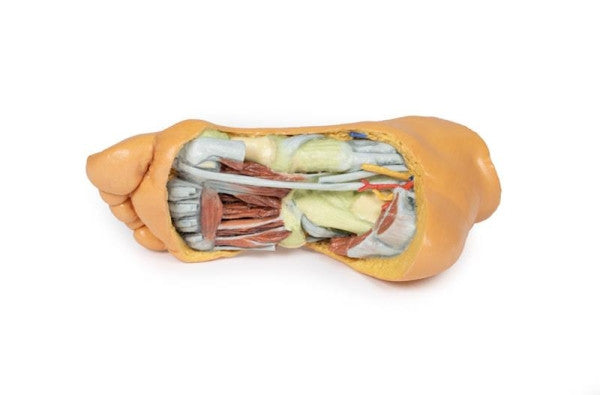
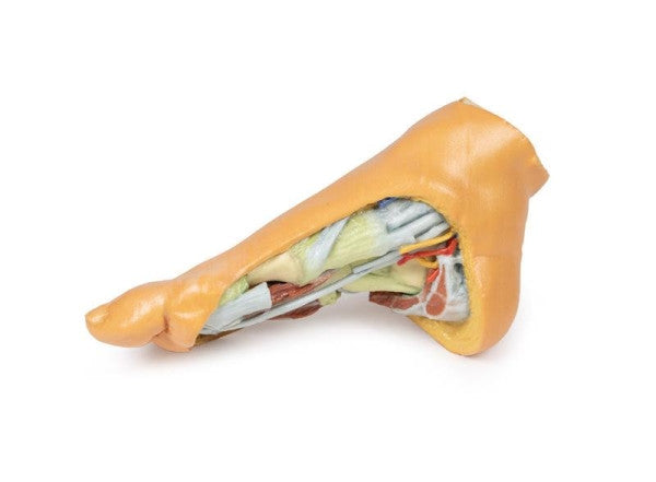
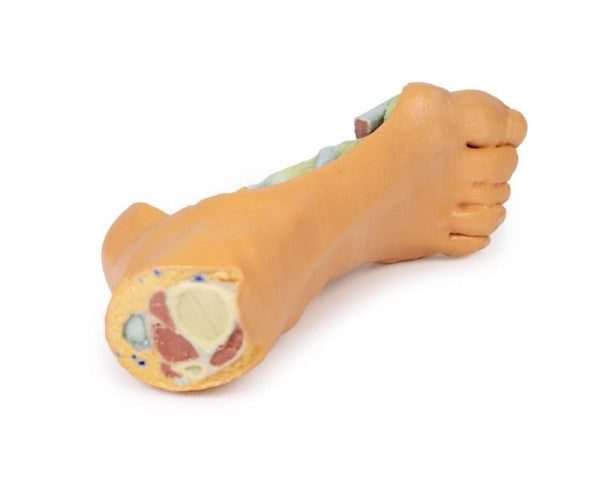
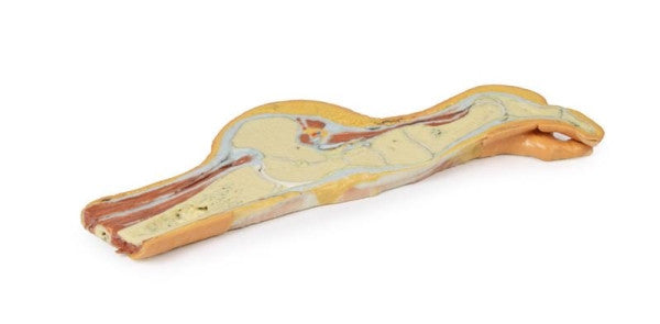
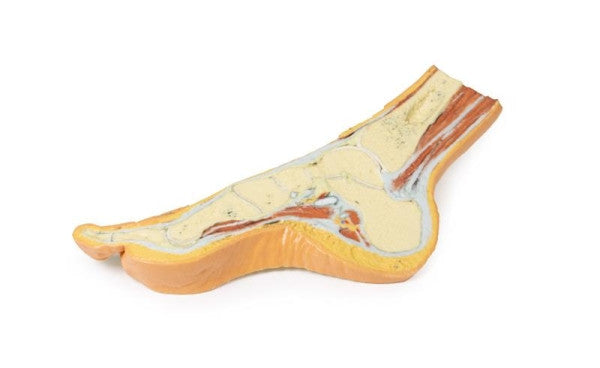
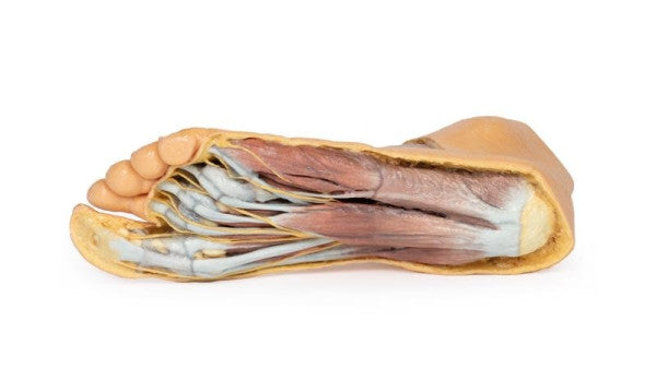
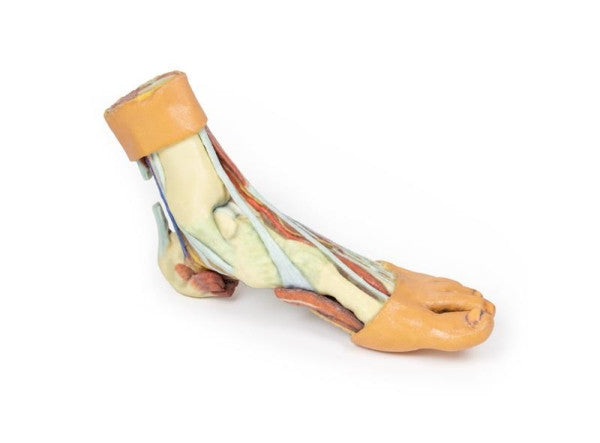
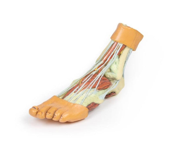
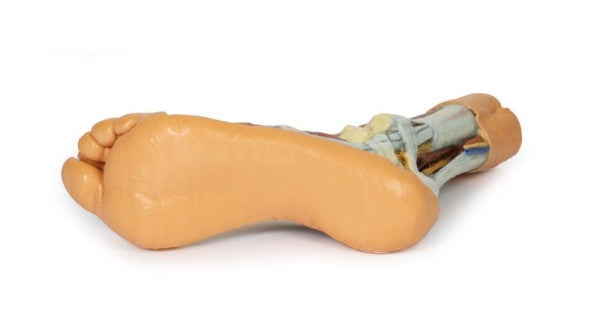
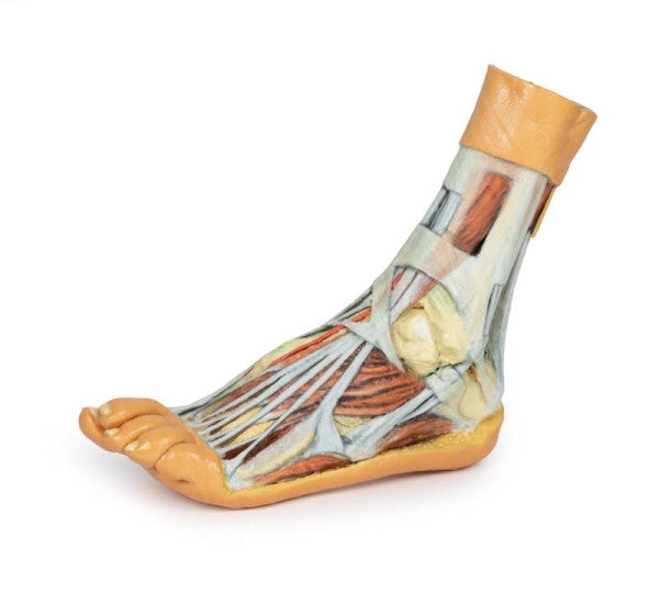
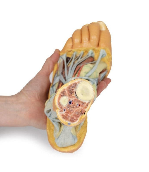
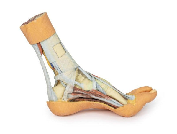
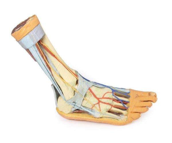
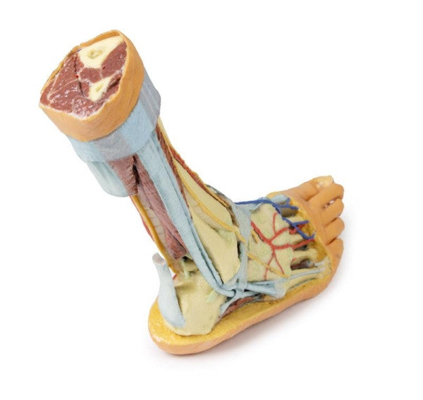
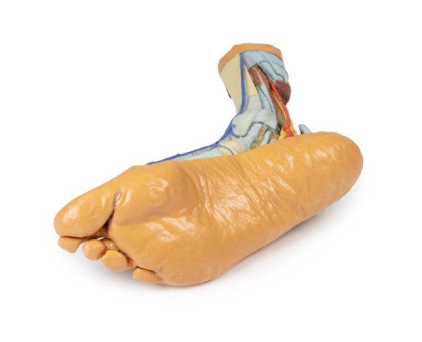
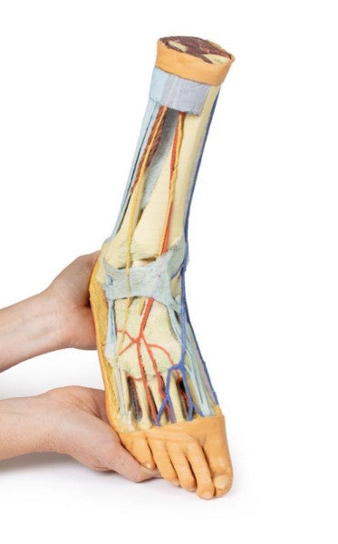
The Foot 3D Printed Anatomy Models showcase various perspectives of lower limb anatomy. One specimen provides a parasagittal cross-section through the medial aspect of the right distal tibia and foot, emphasising the skeletal structures of the medial longitudinal arch. Another presents a view of deep plantar structures in the right foot, revealing the great saphenous vein, plantar arteries, and nerves.
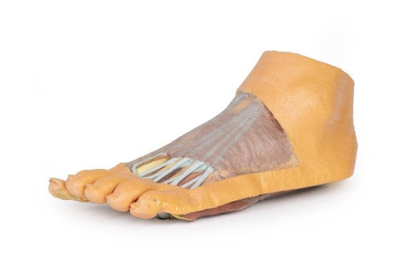
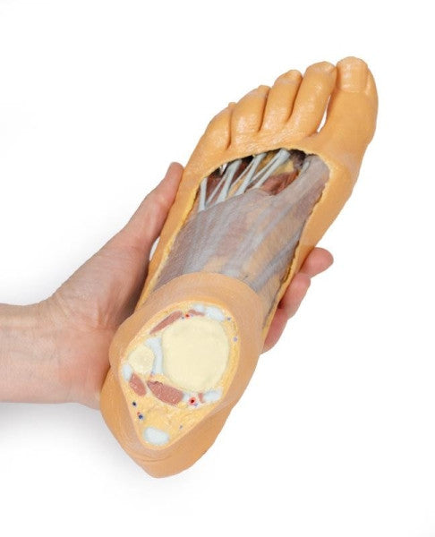
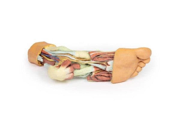
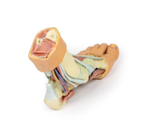
Additional specimens feature detailed anatomy of the distal leg and foot, including both superficial and deep dissections, highlighting muscles, tendons, and neurovascular structures. These models serve as valuable educational tools for studying lower limb anatomy.