


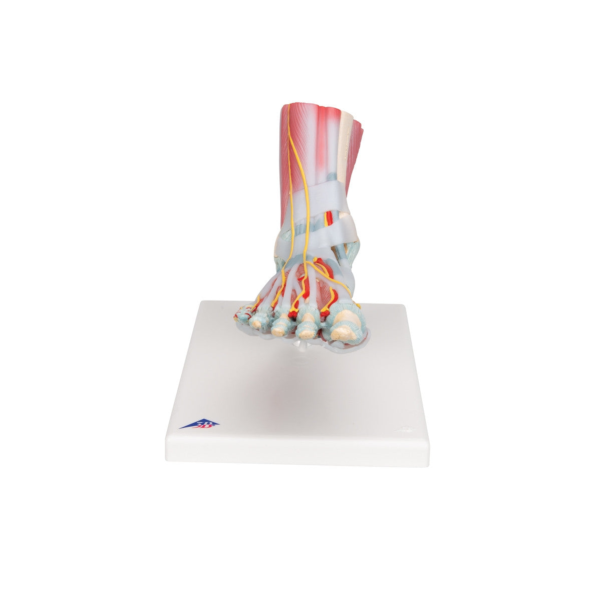
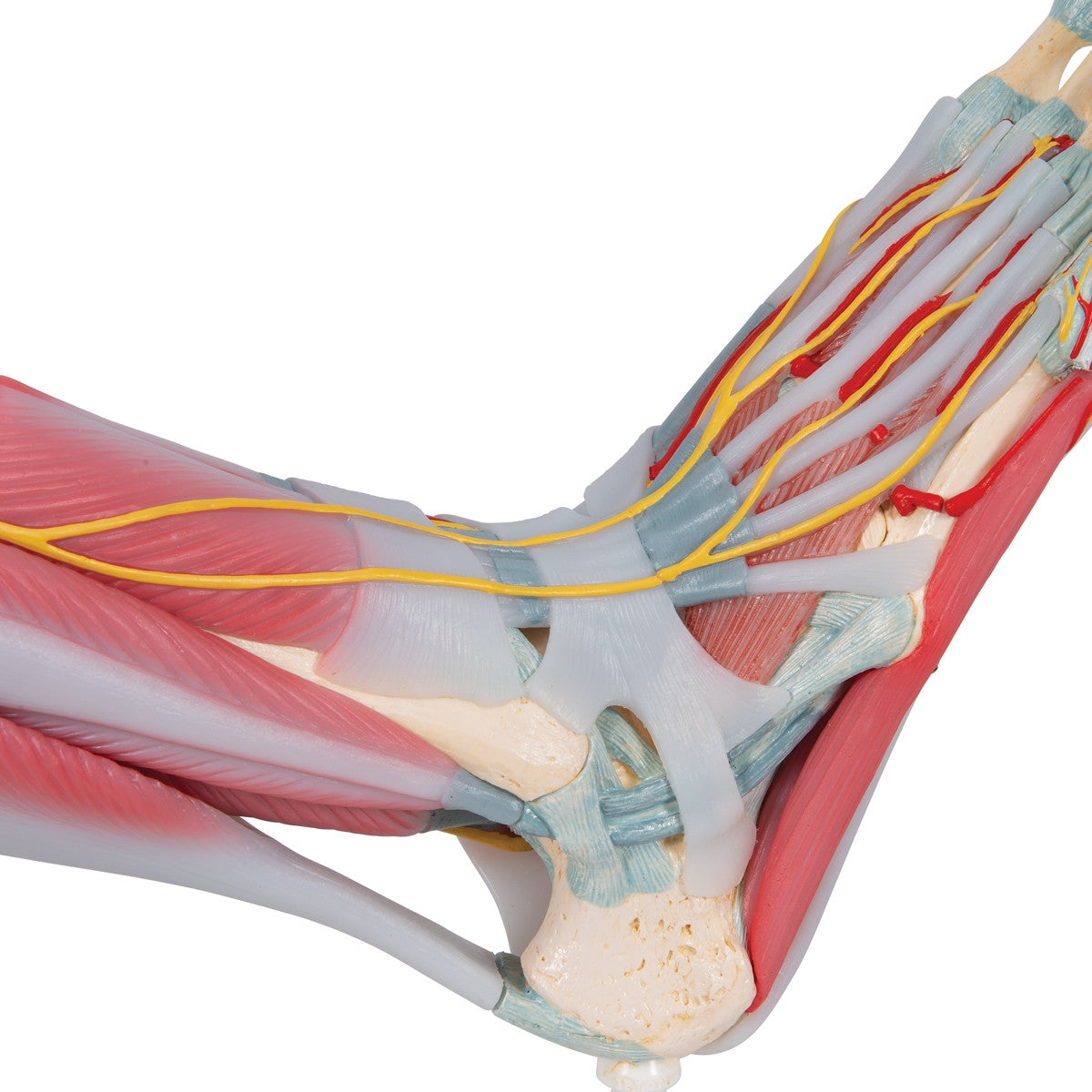
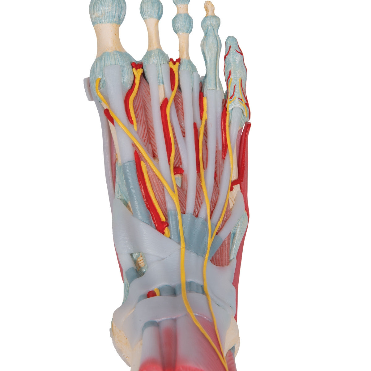
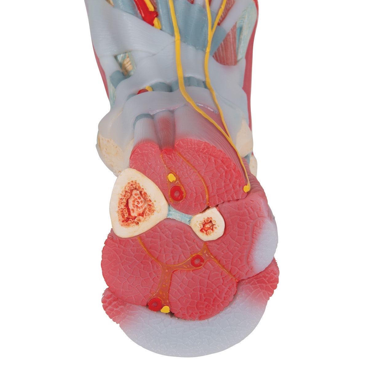
A detailed model of the foot and lower leg showing the bones, muscles, tendons, ligaments, nerves, arteries and veins.
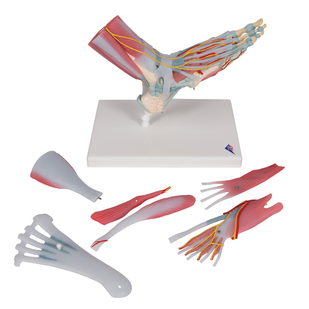
This Foot and Ankle Model with Muscles and Ligaments is an anatomically correct model of the lower leg which can be taken apart into 6 parts for detailed study. Model M34/1 / 1019421 features the foot and ankle bones as well as the major muscles, tendons, ligaments, nerves, arteries and veins of the foot.
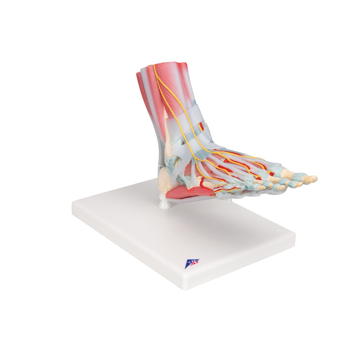
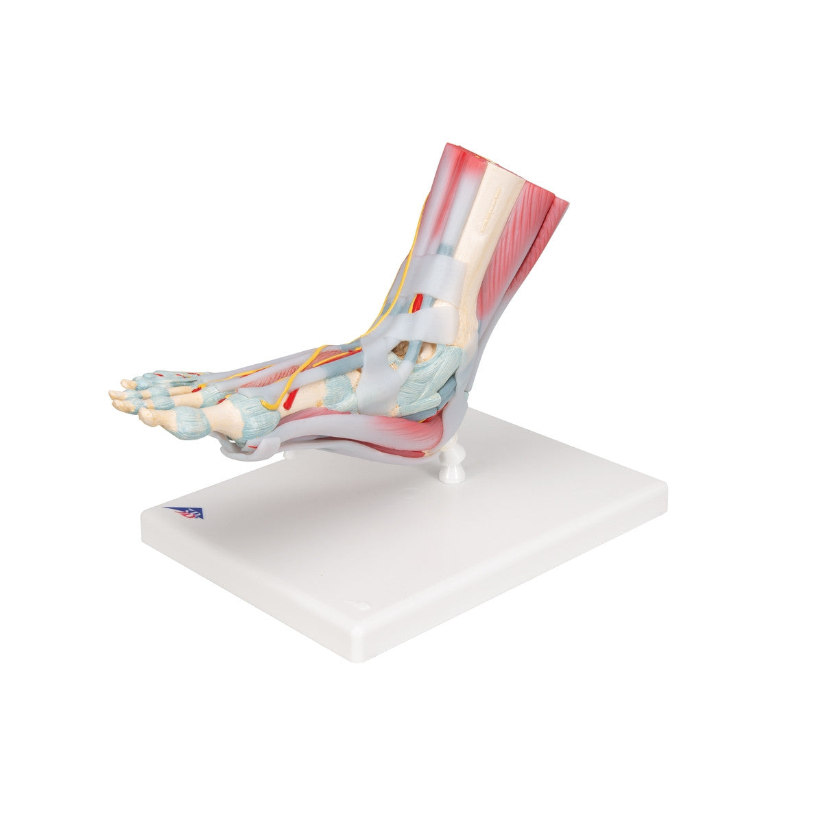
The front part of the foot model features the extensor muscles of the lower leg. The tendons are visible and can be traced under the transverse and crucial crural ligaments to their insertions points, along with the tendon sheaths of the foot area.
The back part of the foot muscle model features a removable gastrocnemius muscle, which when removed shows the deeper anatomical elements. The sole of the foot model is made up of three layers; the first showing the flexor digitorum brevis muscle which can be detached to reveal the quadratus plantae, the tendon of the flexor digitorum longus and the flexor hallucis muscle. This second layer can also be removed for even deeper study of the bones and anatomical features of the foot.
An ideal model for detailed study of the musculature of the foot ideal for universities, hospitals or clinics.
This model comes with 3B Scientific 3B Smart Anatomy app included for FREE. This features access to an anatomy course, including 3 digital anatomy lectures, 117 different virtual anatomy models and 39 anatomy quizzes. It also offers a FREE warranty upgrade from 3 to 5 years with every product registration. To unlock these benefits, scan the label located on your 3B Scientific anatomy model and register online.