


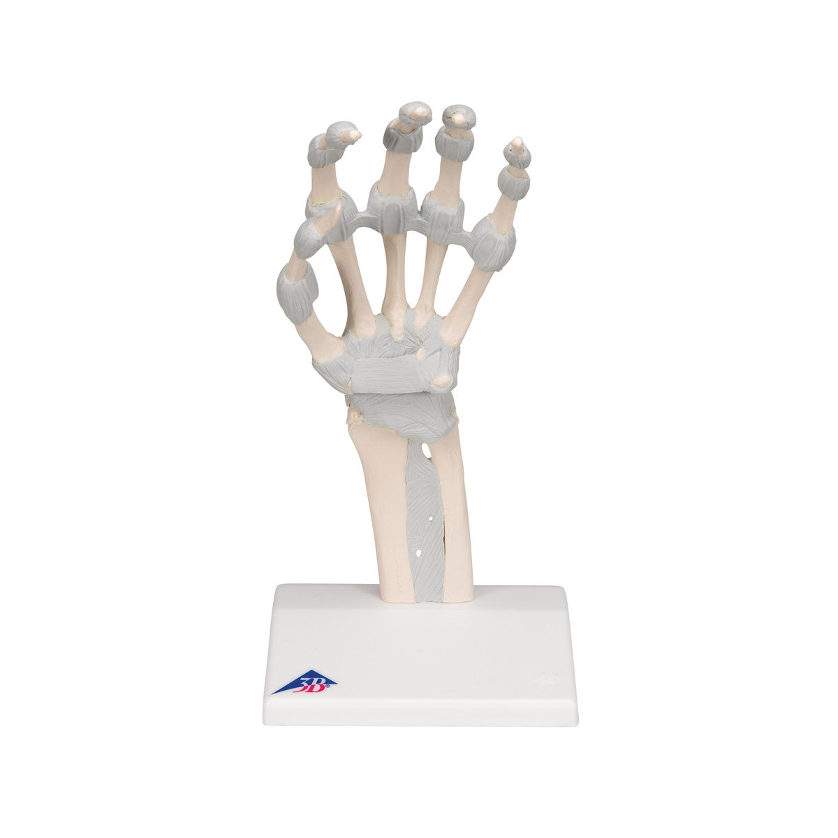
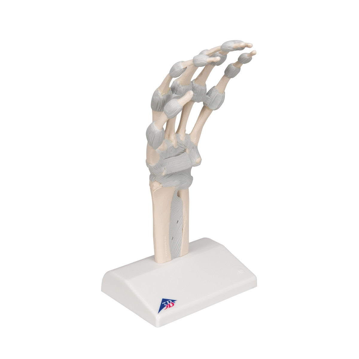
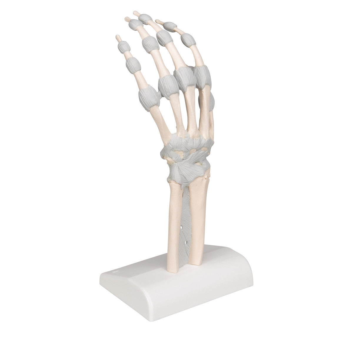
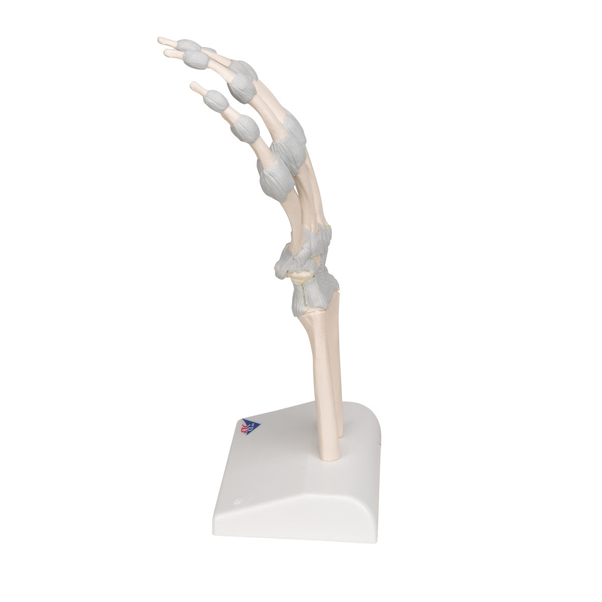
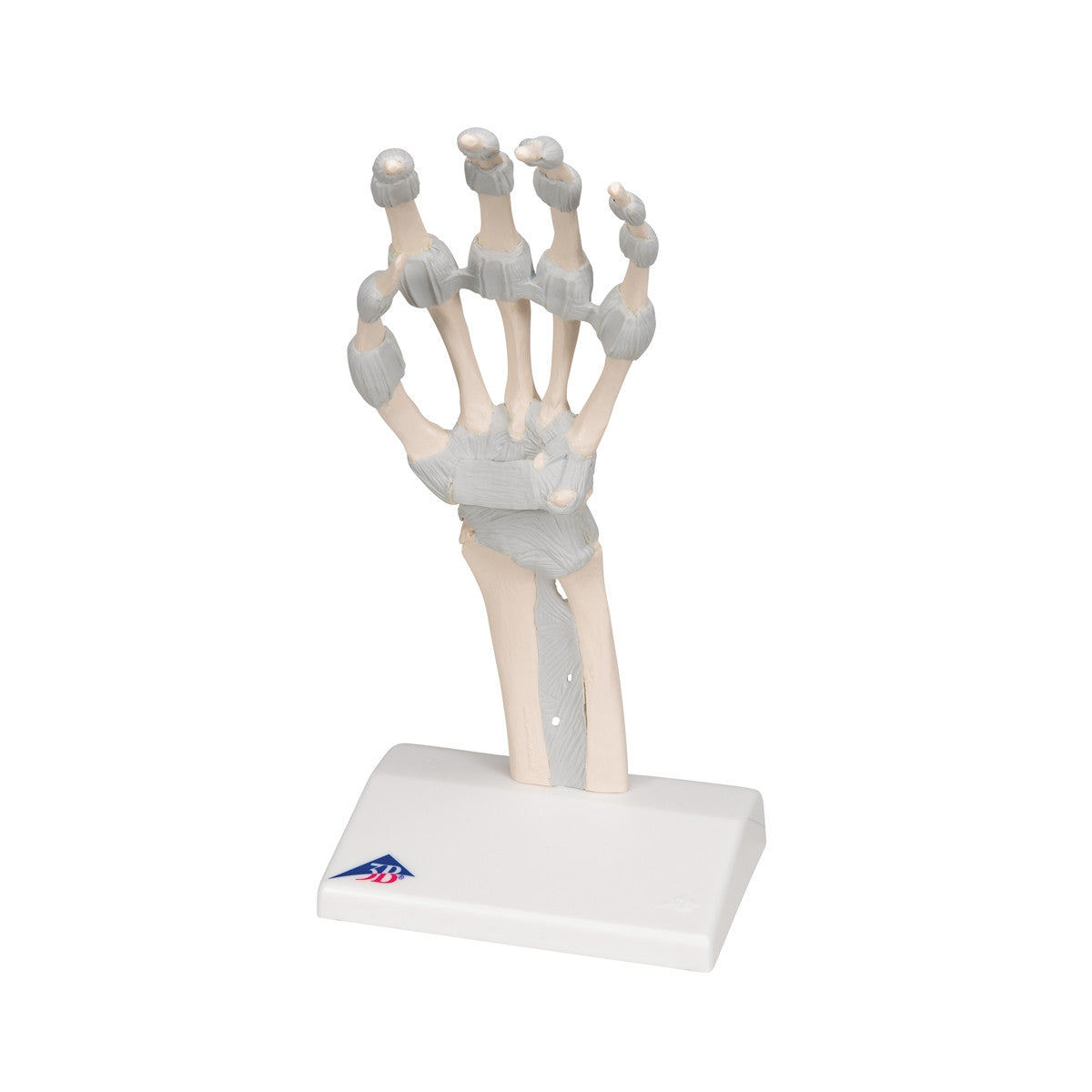
A detailed replica with flexible wrist and hand ligaments to show movement.
This M36 / 1013683 hand skeleton anatomy model accurately shows the ligaments in the human hand. Designed as a teaching aid, the model is perfect for use in anatomy classes, as well as for medical students, physiotherapists and occupational therapists.
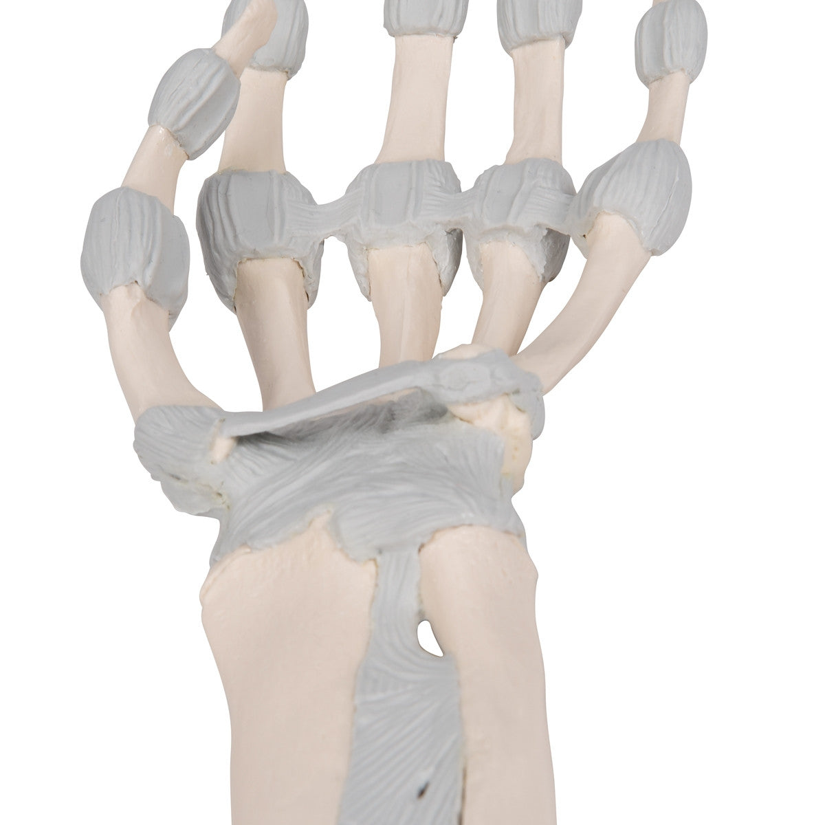
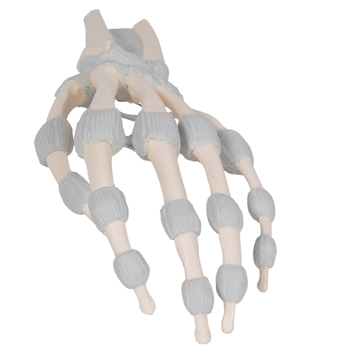
The carpals (ossa carpi), the metacarpals (ossa metacarpi) and finger bones (ossa digitorum manus) are represented as osseous structures. The radius and the ulna can be seen in the distal area of the forearm. The membrana interossea, the fibrous layer of connective tissue, can be seen extended between both of these long bones.
The flexor retinaculum, which encloses and forms the roof of the carpal tunnel is also represented.
The ligaments (membrana interossea and flexor retinaculum) have been recreated flexibly, allowing for the realistic simulation of functional movement, especially in the wrist joints to aid in accurate teaching.
Download Hand Skeleton Model with Elastic Ligaments M36 / 1013683 product manual here.
This model comes with 3B Scientific 3B Smart Anatomy app included for FREE. This features access to an anatomy course, including 3 digital anatomy lectures, 117 different virtual anatomy models and 39 anatomy quizzes. It also offers a FREE warranty upgrade from 3 to 5 years with every product registration. To unlock these benefits, scan the label located on your 3B Scientific anatomy model and register online.