


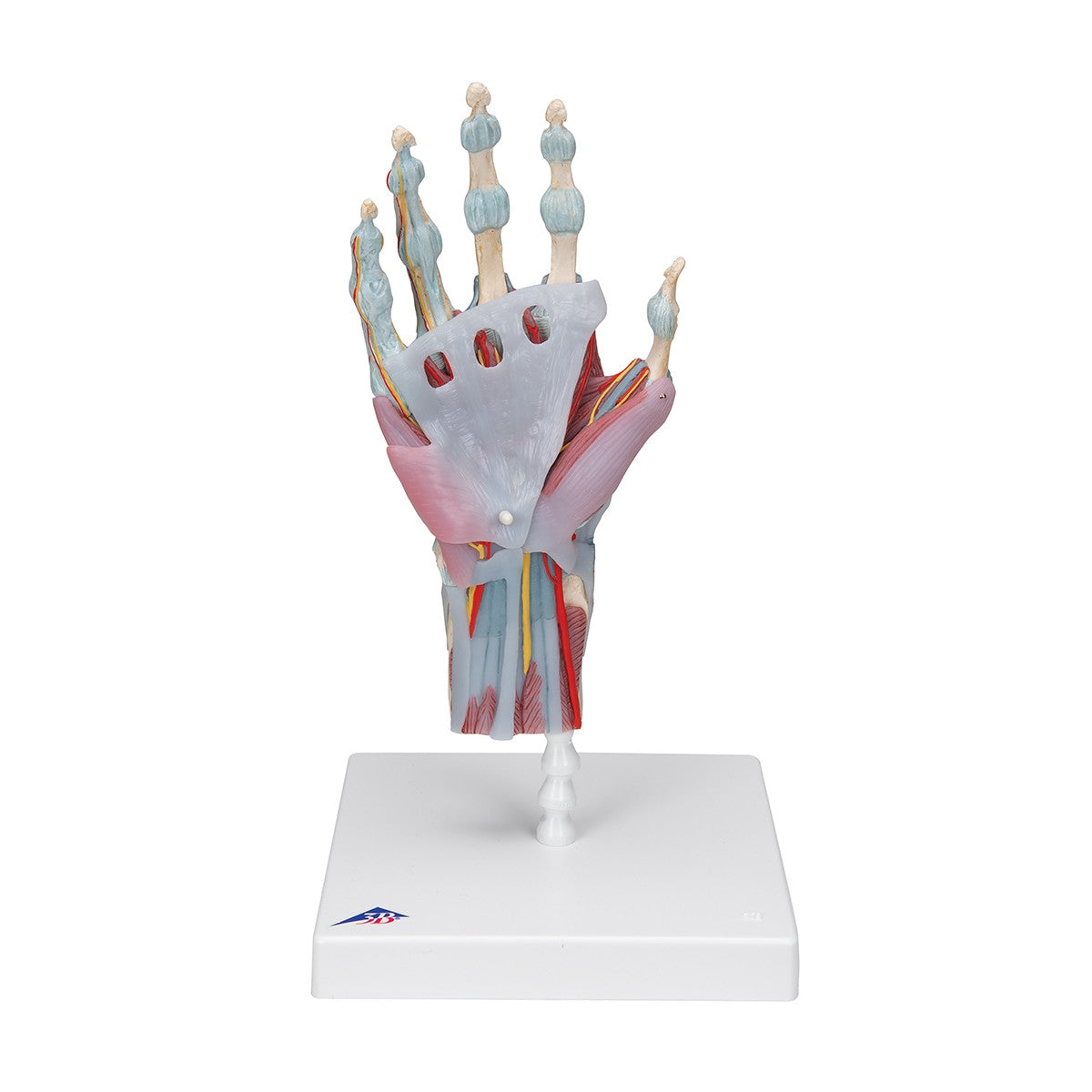
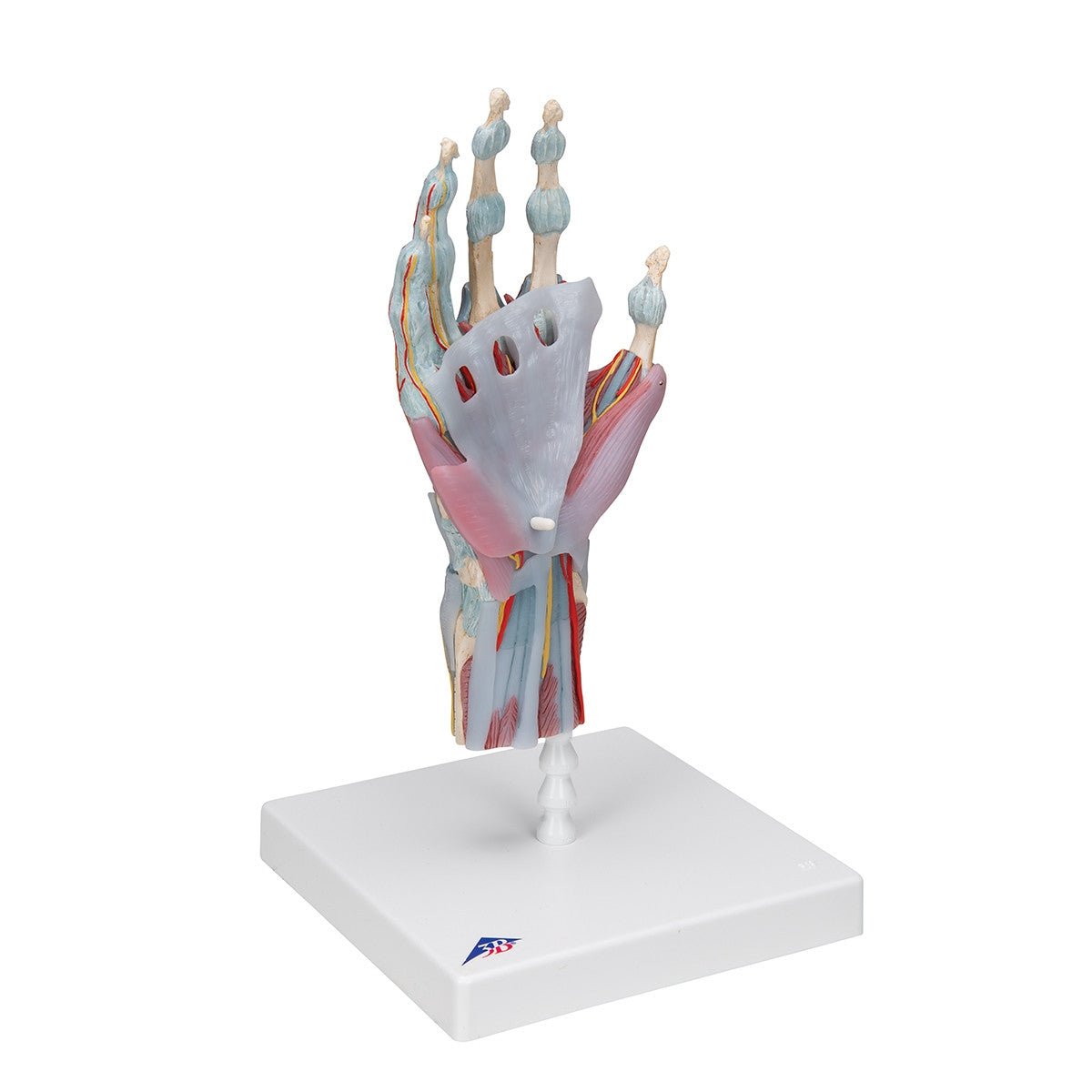
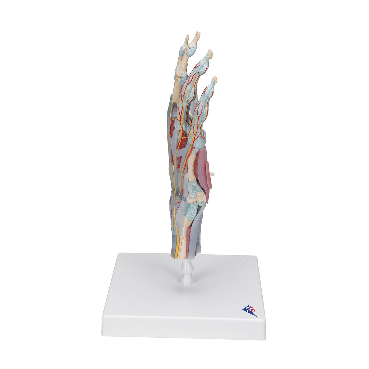
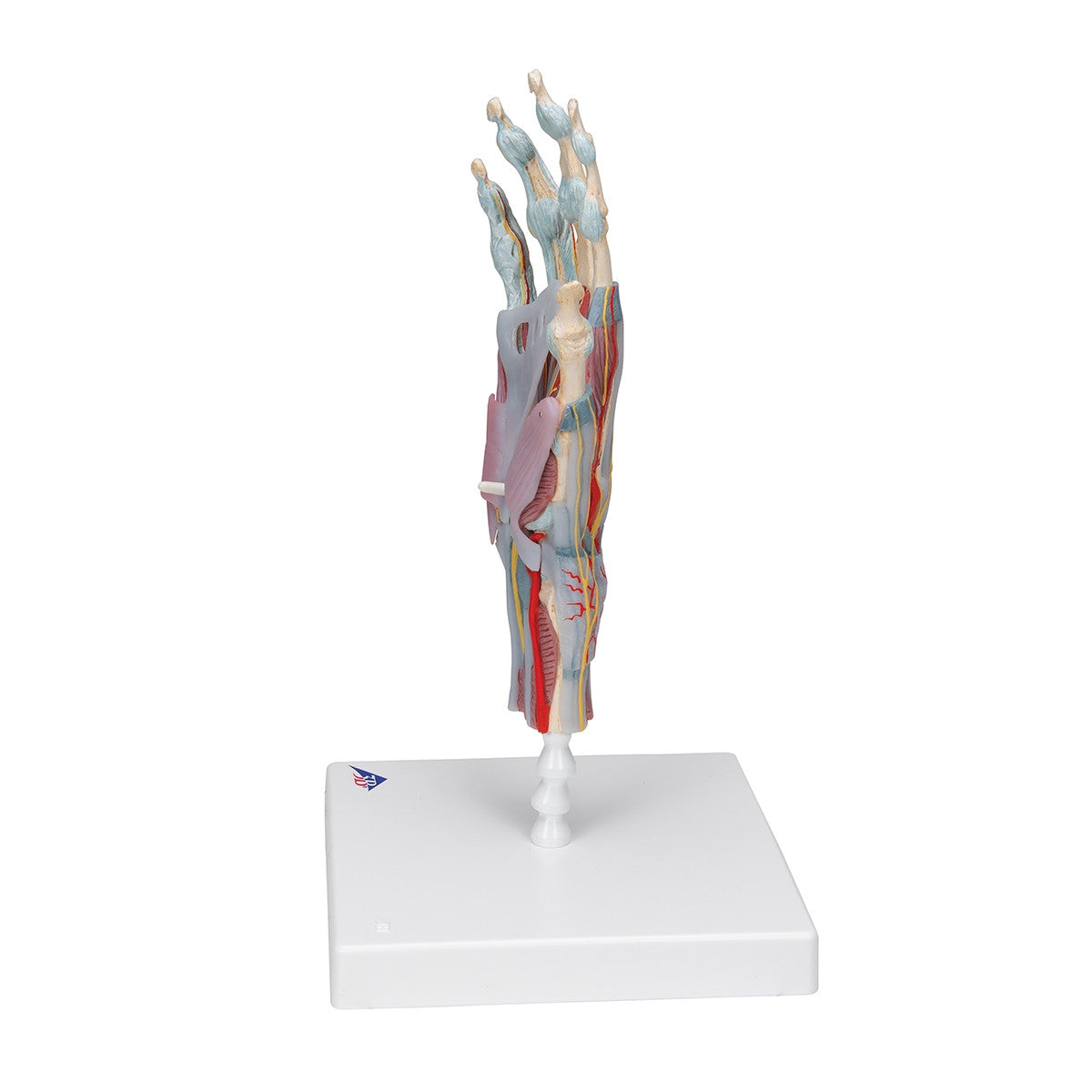
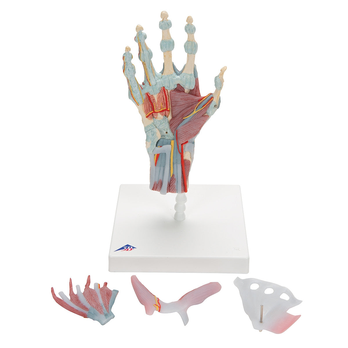
A detailed model of the human hand for study of the ligaments, nerves, blood supply and muscles, including the intrinsic muscles.
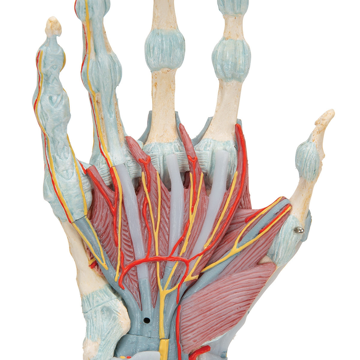
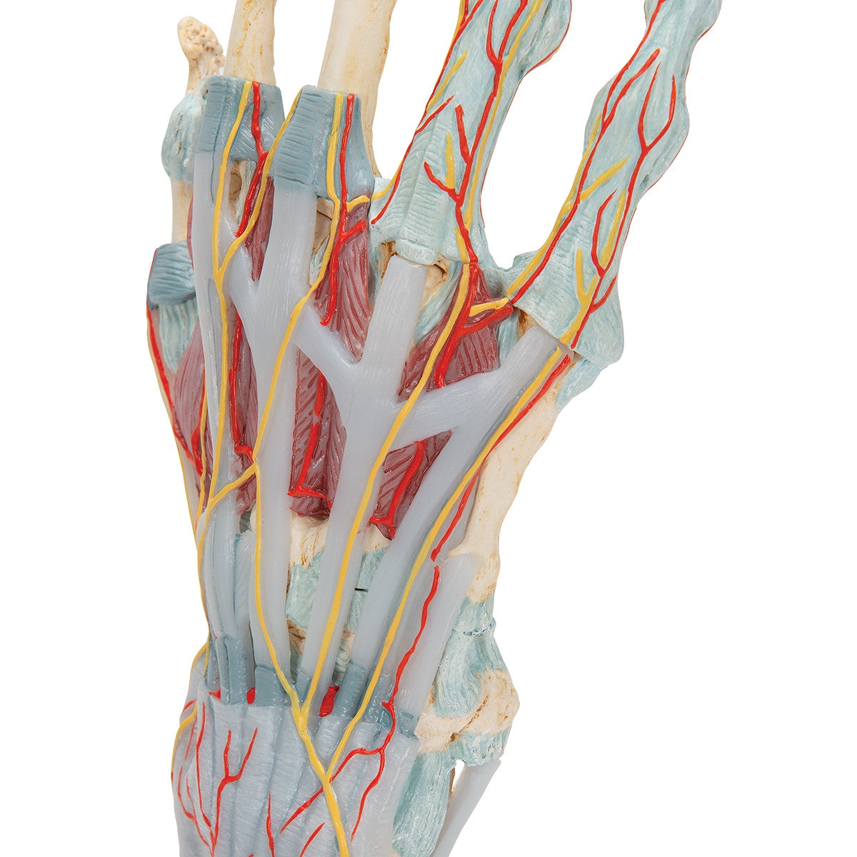
This anatomical Hand Model shows the bones, muscles, tendons, ligaments, nerves, arteries and veins of the human hand and lower forearm. The dorsal side of the model shows the extensor muscles and portions of the tendons at the wrist as they pass under the extensor retunaculum. The palmar face of the hand is made up of three layers, the first two are detachable allowing for detailed study of the deeper anatomical layer of the hand.
The hand model M33/1 / 1000358 also features clinically important structures such as the median nerve and superficial palmar arterial arch can be studied in detail. The deepest anatomical layer allows for study of the intrinsic muscles and deep palmar arterial arch as well as other details of the anatomy of the hand. The Hand Skeleton Model with Ligaments and Muscles is an ideal tool for teaching or studying the complex anatomy of the human hand.
This model comes with 3B Scientific 3B Smart Anatomy app included for FREE. This features access to an anatomy course, including 3 digital anatomy lectures, 117 different virtual anatomy models and 39 anatomy quizzes. It also offers a FREE warranty upgrade from 3 to 5 years with every product registration. To unlock these benefits, scan the label located on your 3B Scientific anatomy model and register online.