


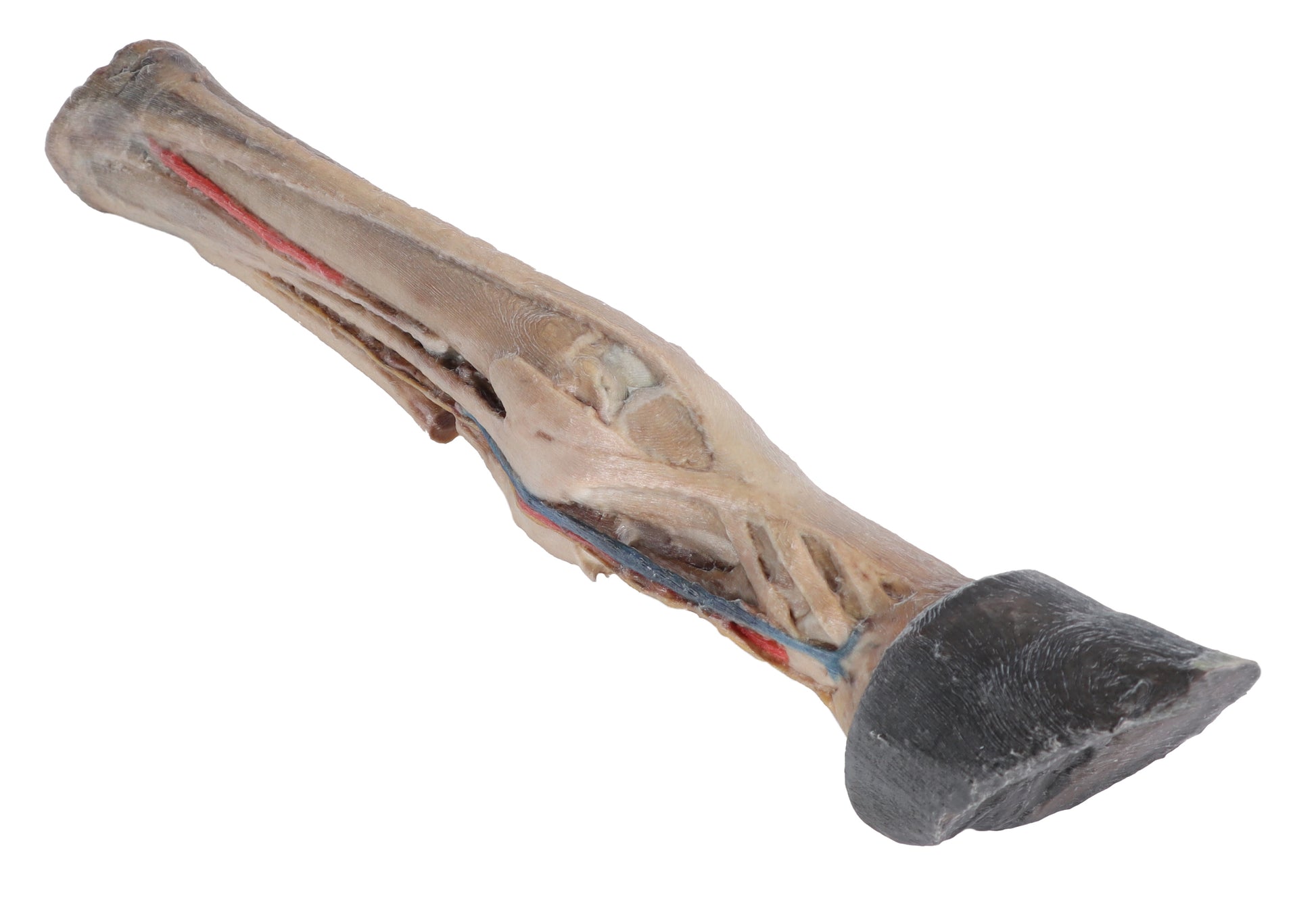
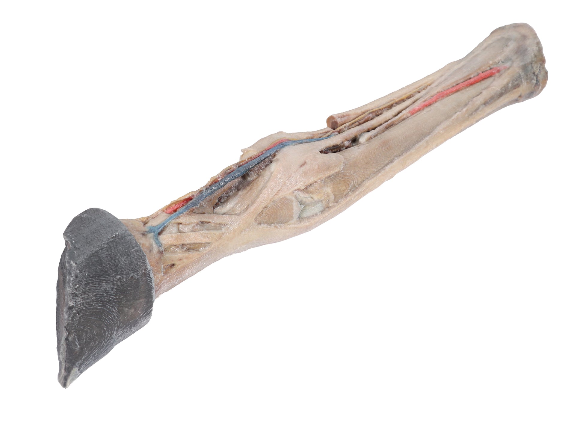
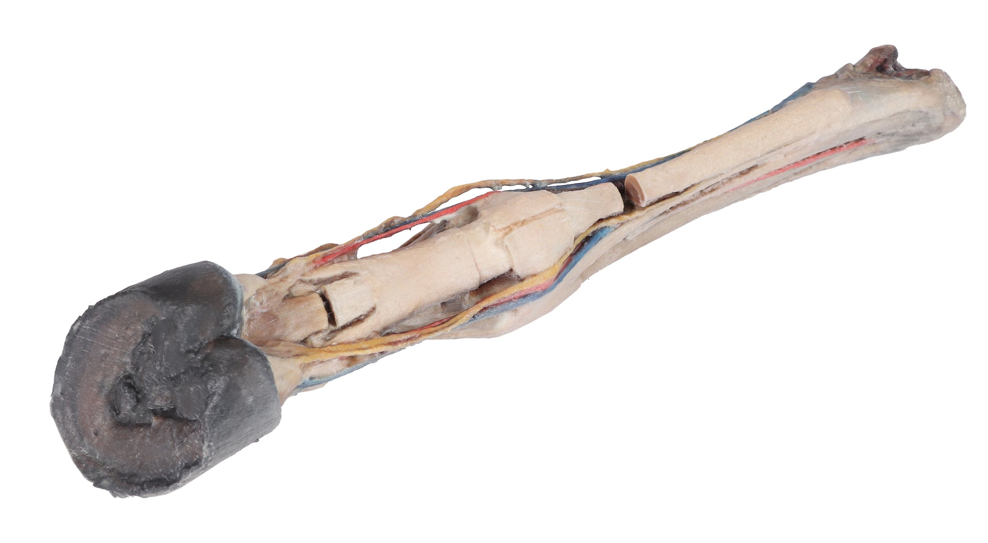
This 3D Printed Horse Foot Model provides a comprehensive view of the anatomical relationships between vascular, nervous, ligamentous, and tendinous structures.
On the proximal dorsal aspect, the two digital extensor tendons are clearly visible. The plantar digital neurovascular bundles are located on either side of the flexor tendons. The metatarsophalangeal and proximal interphalangeal joints are fully exposed, displaying all associated ligaments.
Positioned between the plantar surface of the metatarsal bones and the digital flexor tendons, the interosseous ligament is shown along with its sesamoid and extensor branches.
This model is part of a range of detailed 3D printed veterinary models, exclusively available in the UK via AnatomyStuff, ideal for advanced anatomical study.