


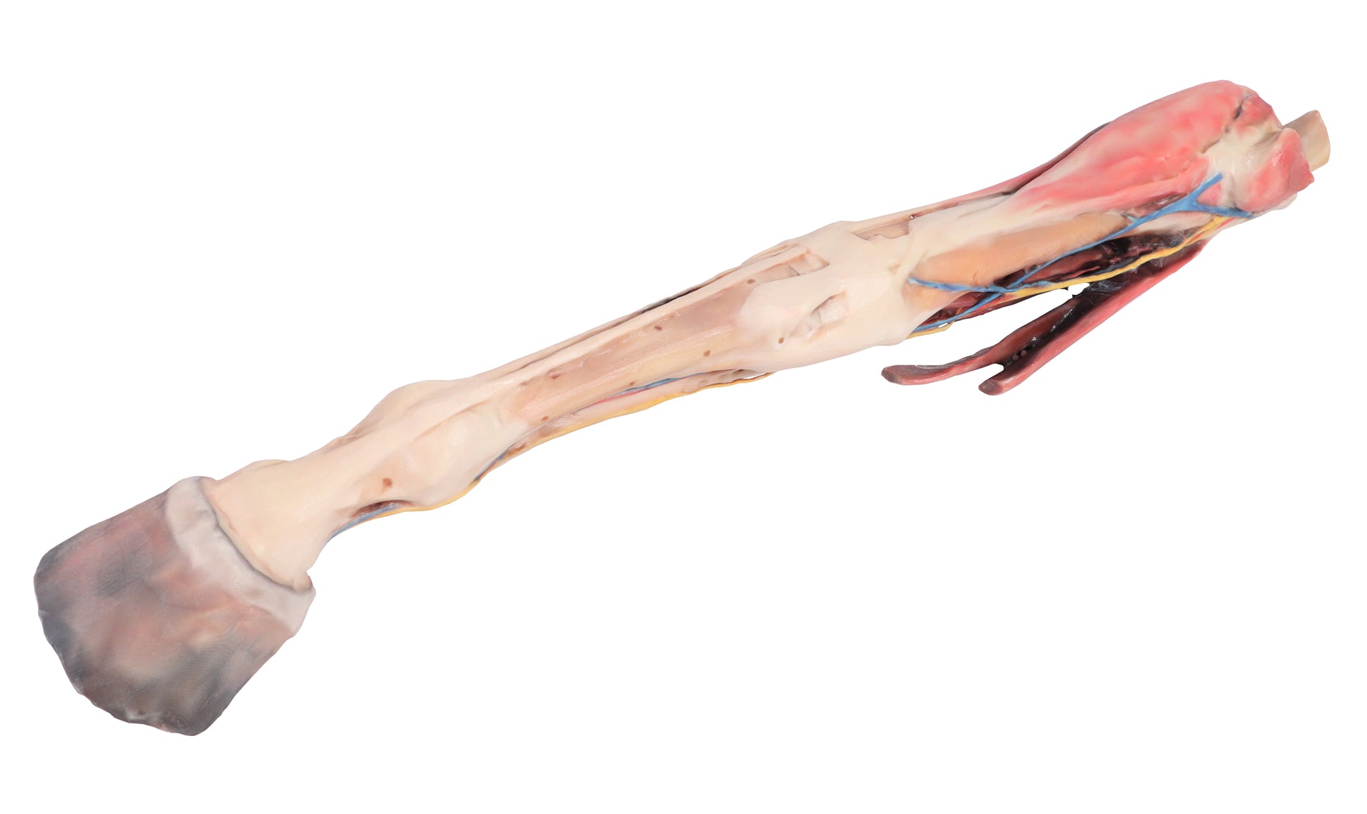
The 3D Printed Equine Fore Limb Model displays the superficial anatomy of the right forelimb, extending from the elbow to the hoof.
It highlights the carpus and the digital extensor and flexor muscles in relation to the median and ulnar nerves. Major branches of the median artery, along with the median and cephalic veins, are preserved. Below the carpus, the palmar nerves are shown in relation to the digital flexor tendons, and distal to the metacarpophalangeal joint, the palmar digital neurovascular bundles are visible on both sides
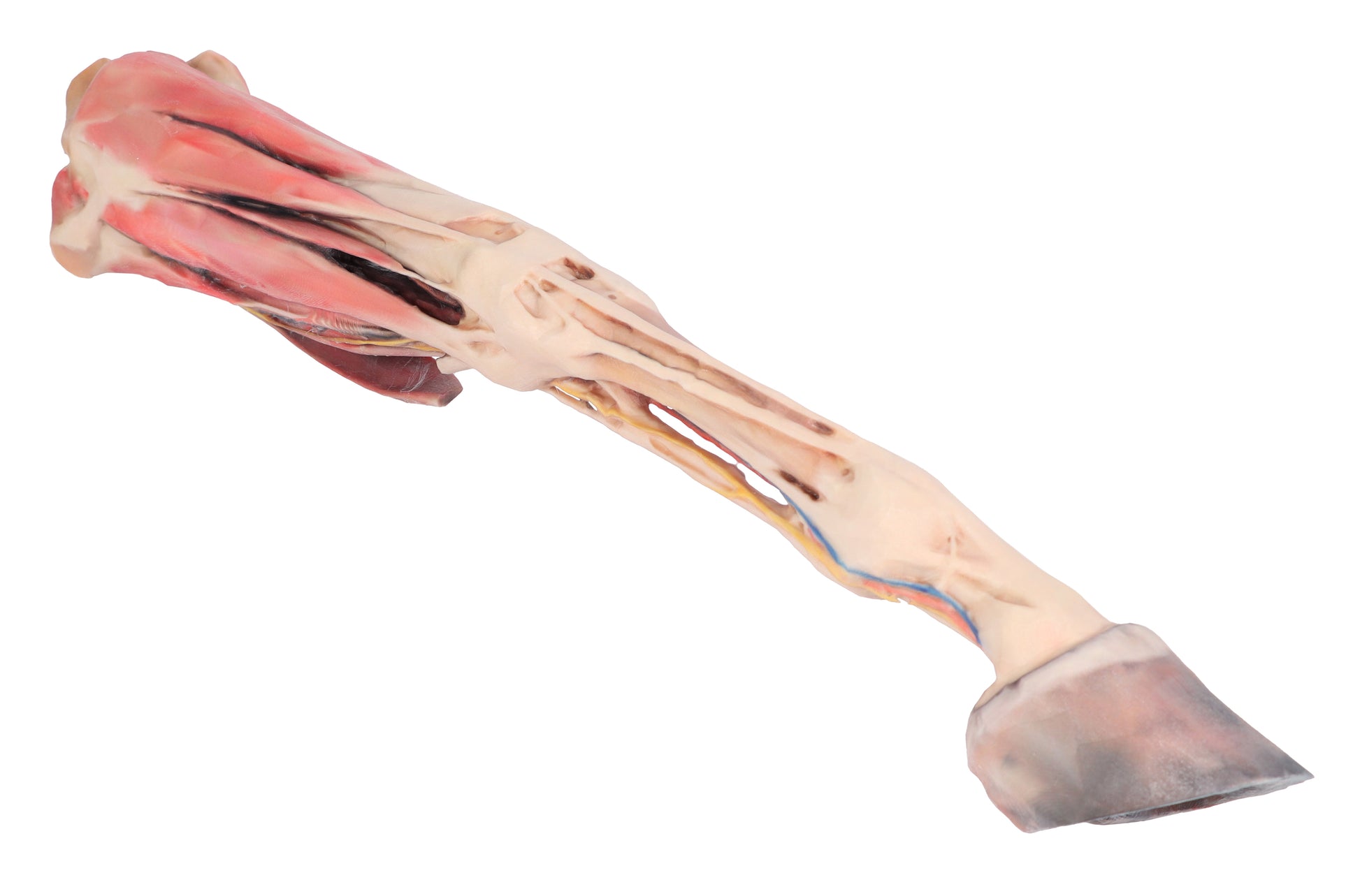
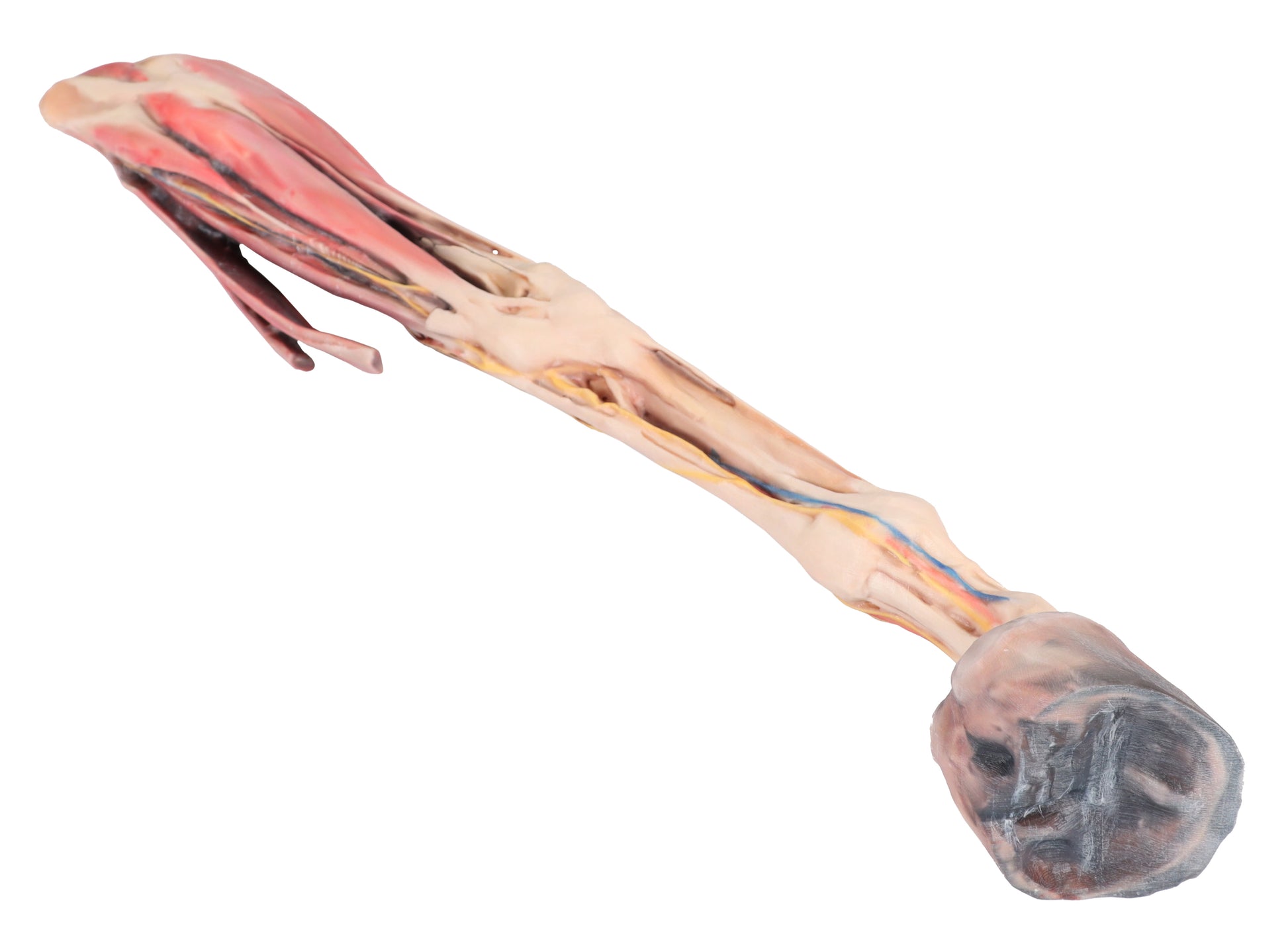
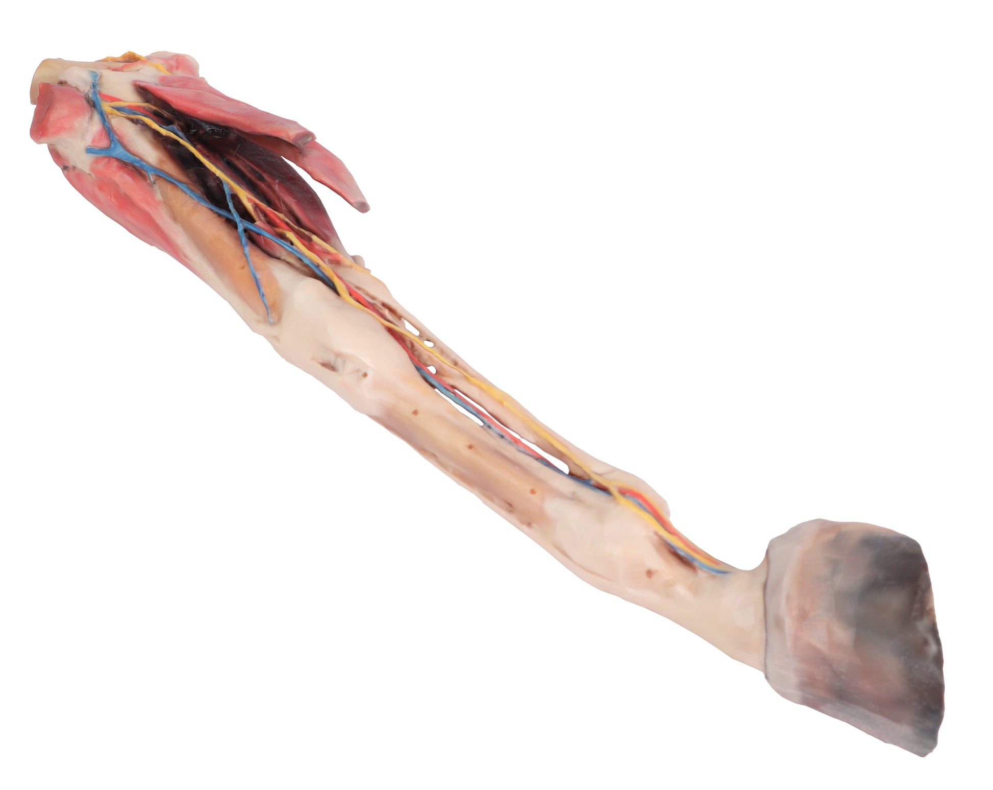
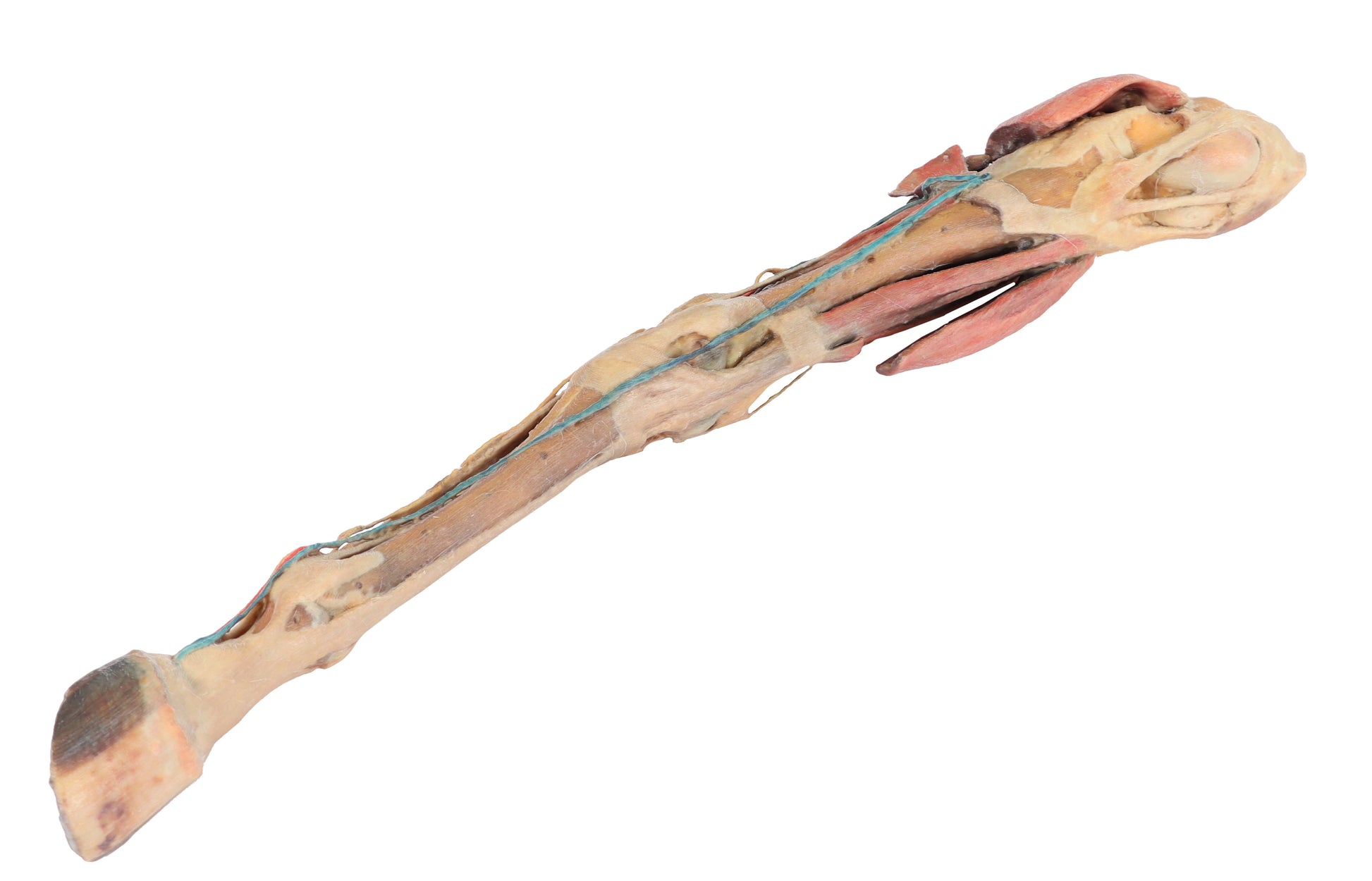
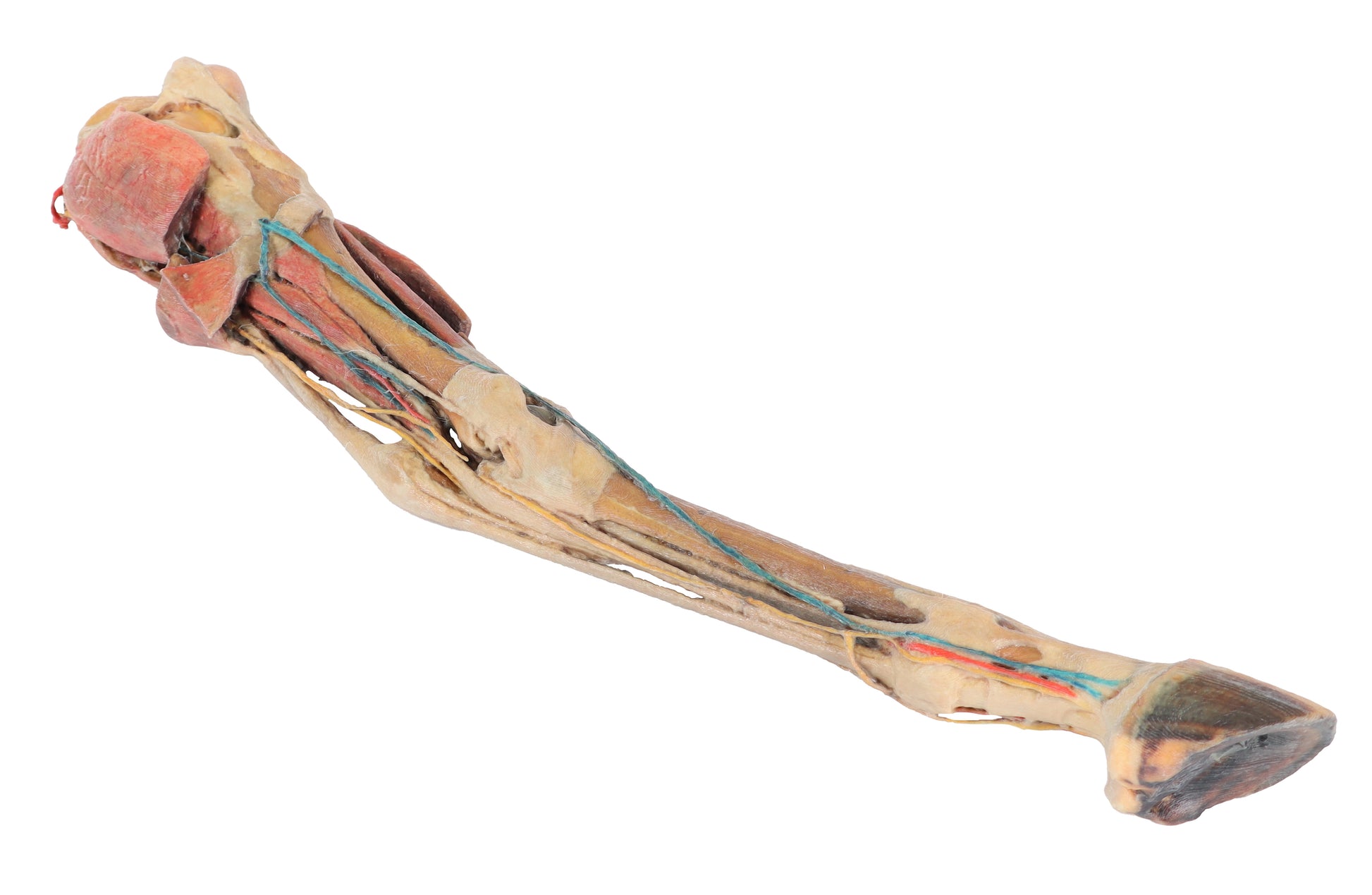
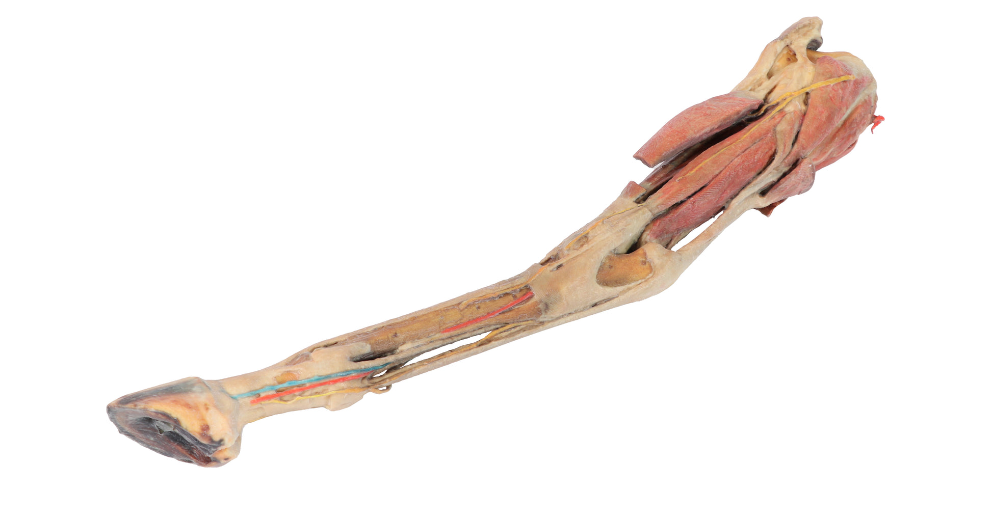
The 3D Printed Equine Horse Hind Limb Model presents a superficial dissection of the lower limb, extending from the mid-thigh to the hoof.
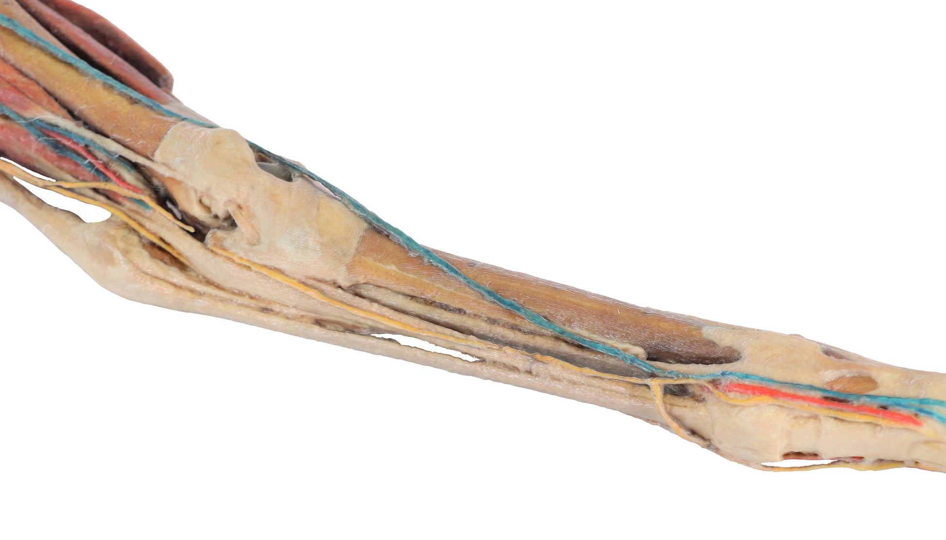
The extensor and flexor muscles of the stifle, tarsus, and foot joints are dissected from their origins on the femur and tibia to their insertions on the tarsal, metatarsal, and phalangeal bones.
The joint capsules of the stifle and tarsus have been opened to expose the menisci, major ligaments, and tendon topography. Nerves and blood vessels are also dissected and visible in the popliteal fossa, tarsal, and digital regions.
This model is part of a range of detailed 3D printed veterinary models, exclusively available in the UK via AnatomyStuff, ideal for advanced anatomical study.