Trusted Supplierto the NHS
Puchase OrdersAccepted
Shop Over5,000 Products
Free UK DeliveryOver £20
Over 100,000Happy Customers
Exc. VAT
Regular price £372.00
Inc. VAT

SKU : MP2091
Select Model:

Delivery: 4 - 6 weeks

Subtotal: £372.00

Renal Pathology 3D Printed Anatomy Model

Brand: Erler Zimmer | Made in Germany

<strong>We Accept Purchase Orders</strong>
We Accept Purchase Orders

Send your purchase order directly to us. Email orders@anatomystuff.co.uk

<strong>Price Match Service</strong>
Price Match Service

Should you find the same item at a lower price elsewhere, please contact us.

Warranty Information

This product comes with a 3 Year warranty

Limited Time Offer:
0 Translation missing: en.additions.countdown_timer.days_label
0 Translation missing: en.additions.countdown_timer.hours_label
0 Translation missing: en.additions.countdown_timer.min_label
0 Translation missing: en.additions.countdown_timer.sec_label

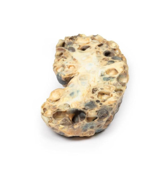
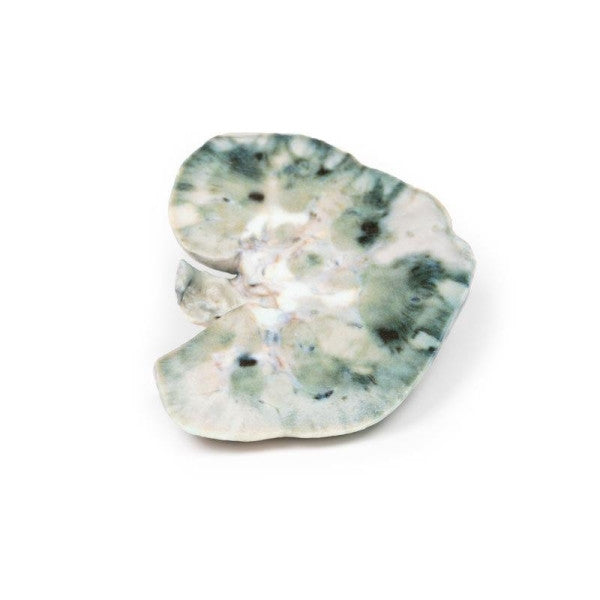
Adult Polycystic Kidney Disease

This enlarged kidney exhibits numerous cysts, up to 3cm in diameter, replacing much of the renal tissue. These translucent-walled cysts, some with varied-coloured contents, created a marbled appearance on the kidney's lobulated external surface. The remaining renal tissue is significantly atrophied due to cyst-induced pressure. This exemplifies adult polycystic kidney disease.
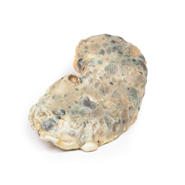
Horseshoe Kidney

The 12cm kidney has a horseshoe shape due to fusion at the lower pole. Ureters emerge anteriorly, and the kidney, bisected horizontally, shows persistent foetal lobulation. The renal pelvis is antero-medially positioned, with ureters running anterior to the fused lower poles.

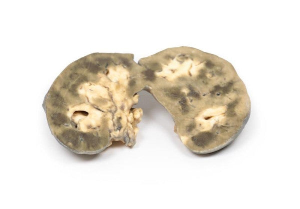
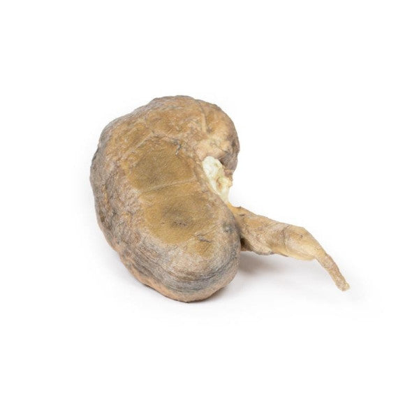
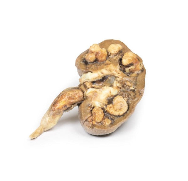
Multiple Renal Calculi

The kidney specimen exhibits noticeable dilatation of the pelvicalyceal system. Extensive renal tissue atrophy is evident, reduced to a thin rim in some areas. A sizable brown-white mottled calculus is present in the pelvis, and a smaller calculus obstructs the ureter lumen. Proximal to the impacted calculus, the ureter shows dilation. Multiple calculi are observable within the calyces of the specimen.
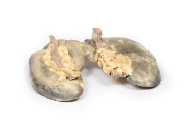
Pyonephrosis

This left nephrectomy specimen exhibits pelvis and calyces dilation with yellow pus remnants. Renal parenchyma displays significant fibrosis, and haemorrhagic necrotic areas near the lateral border, accompanied by two smaller similar areas on the capsular surface. These lesions are likely connected, possibly caused by haemorrhage into an abscess cavity, suggesting a perinephric abscess.

Septic Renal Infarct

The kidney specimen reveals distinct wedge-shaped pale regions in the cortex, extending along renal columns with the apex towards the medulla. The largest is situated laterally in the upper pole, representing infarcted renal tissue. Additionally, there are irregular dark areas indicated of haemorrhage.

Technical Information

Renal Pathology 3D Printed Anatomy Model - MP2091-92-95-96-98 Dimensions: 35.5 cm x 25.5 cm x 14.5 cm Weight: 0.5 kg Manufacturer: Erler Zimmer Made in Germany

Delivery

AnatomyStuff deliver to UK mainland addresses (excluding Northern Ireland and Scottish Highlands and Islands) is FREE for all orders over £20inc VAT.

Delivery to UK mainland addresses (excluding Northern Ireland and Scottish Highlands and Islands) is £6.00 for orders under £20inc VAT.

For delivery to non-mainland UK and European destinations, please contact us for a postage quote.

Please see the delivery estimate in the product information above for an indication of UK delivery times.

For oversized or special order products, an additional shipping and/or restocking charge may apply, however, we will inform you if this is the case before the fulfilment of the order.

Returns Policy

AnatomyStuff's hassle-free return policy guarantees your satisfaction. If you are unhappy with your purchase for any reason you can return it to us in an unused condition within 14 days for a full refund. A return authorisation number is required for all returns, please contact us for this information if you would like to make a return.

Please note: The products featured on our website are designed solely for medical training and educational purposes.