


Life-size kidney model that demonstrates the anatomy and function of kidney stones (nephrolithiasis) and urinary stones (urolithiasis).
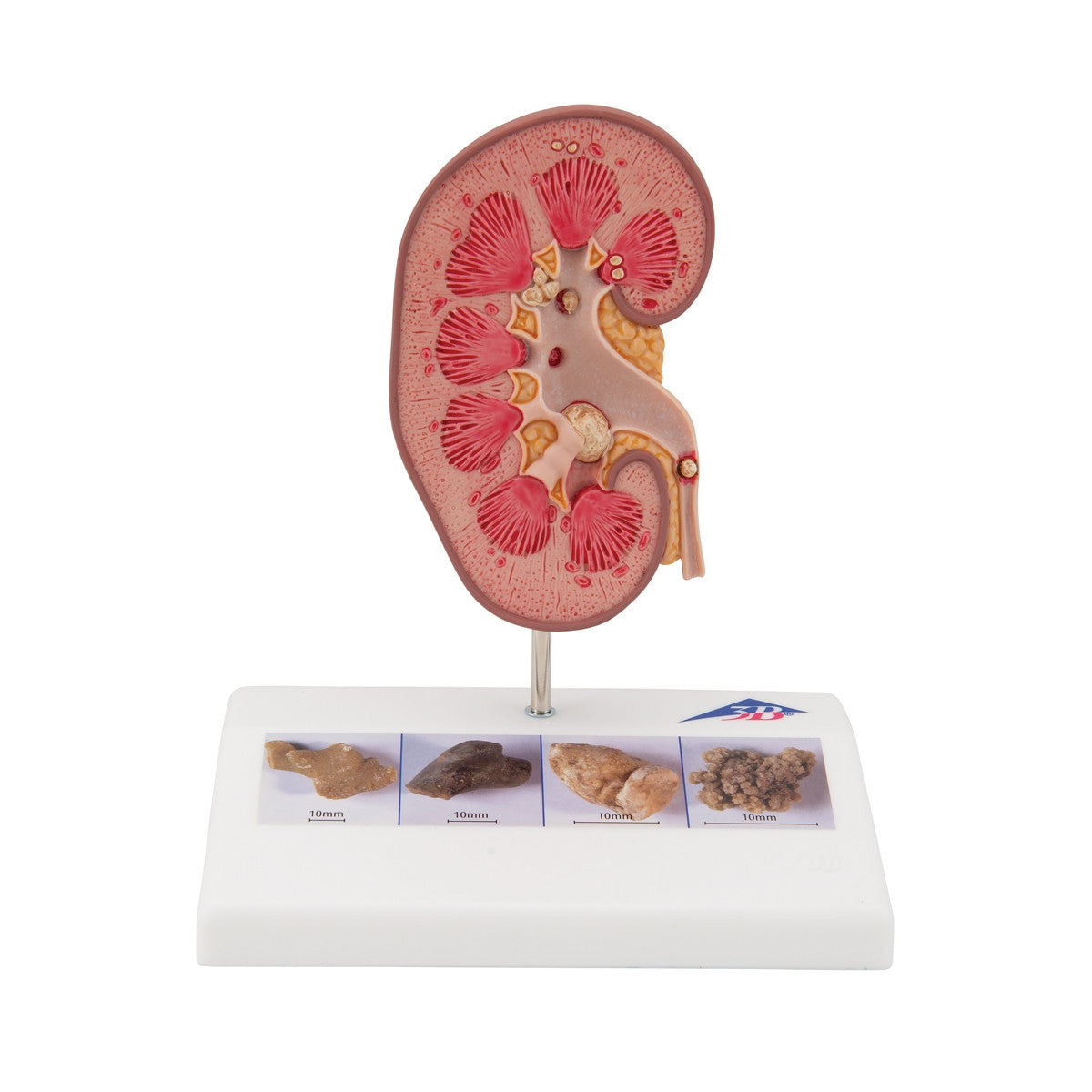
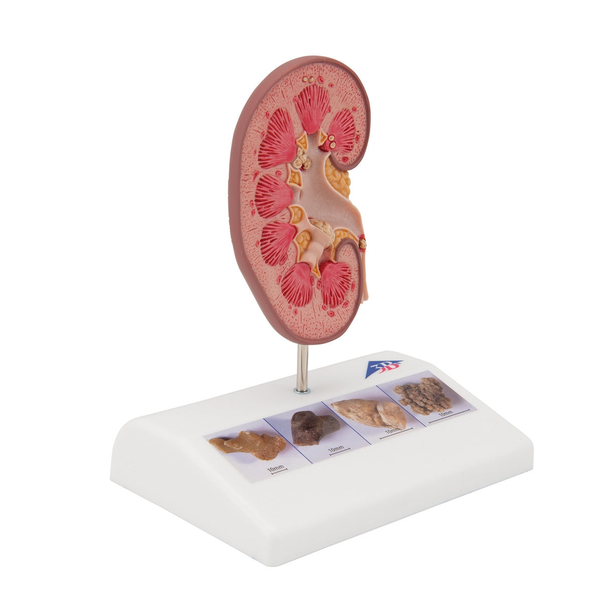
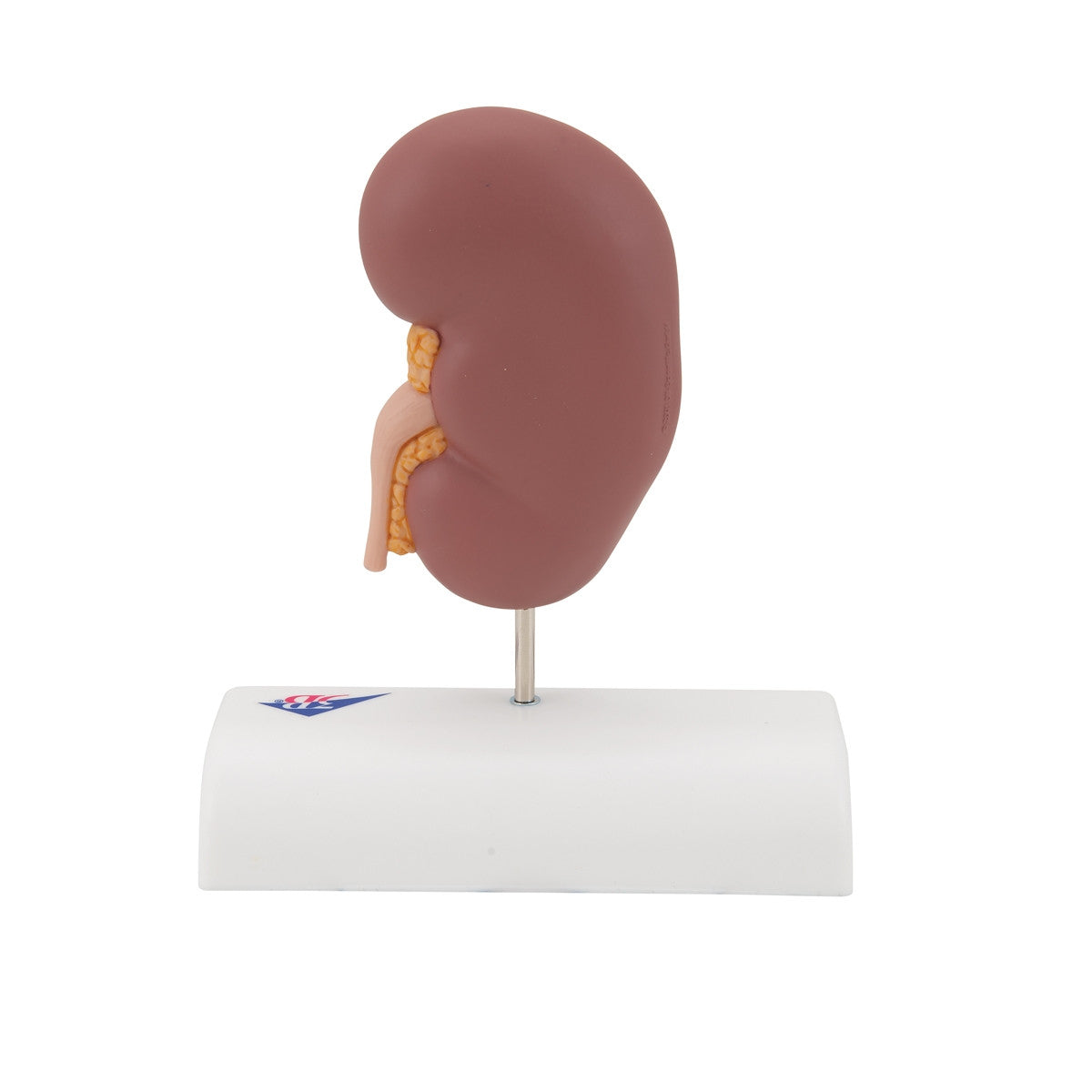
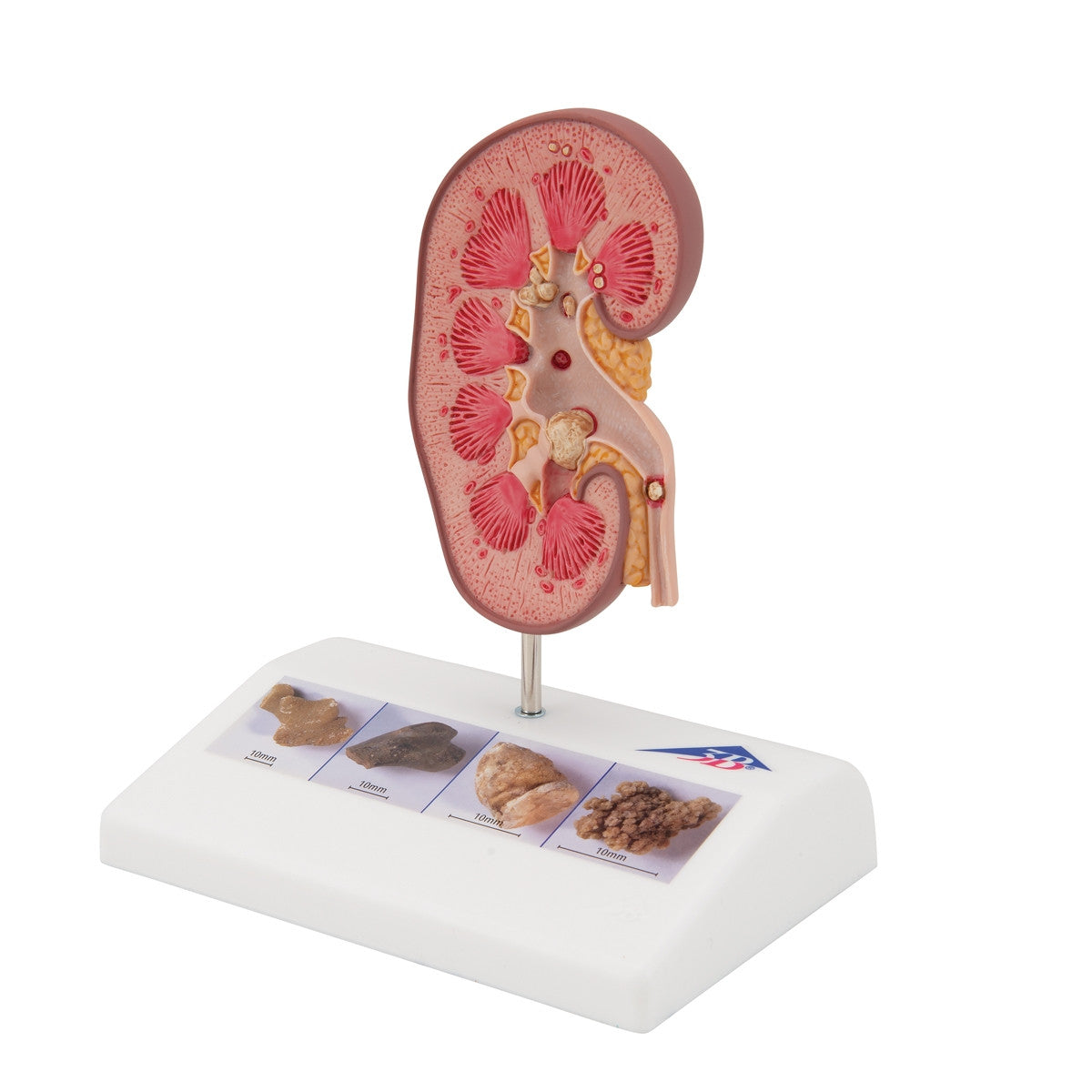
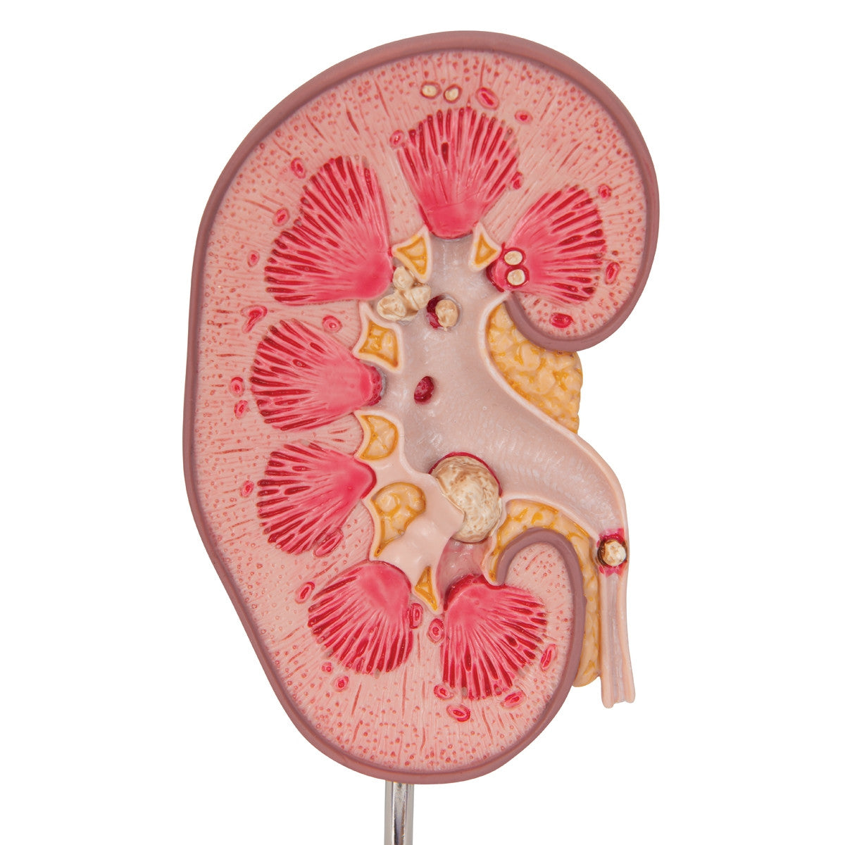
The kidney stone model can be used to demonstrate to patients the anatomy and function of kidney stones (nephrolithiasis) and urinary stones (urolithiasis). Shown in the kidney stone model K29 / 1000316 is an opened right kidney in natural size. The renal calices, the renal pelvis and the ureter are opened as well on the anatomy model so that concretions or stones can be identified in the following typical positions within the kidney:
- In the area of the renal pyramids of the kidney
- In the area of origin of the upper calix group
- In the renal cortex of the kidney
- In the connecting tubule of the lower calix group, causing congestion of the minor calices (partially closed, partially opened) in the kidney in the ureter
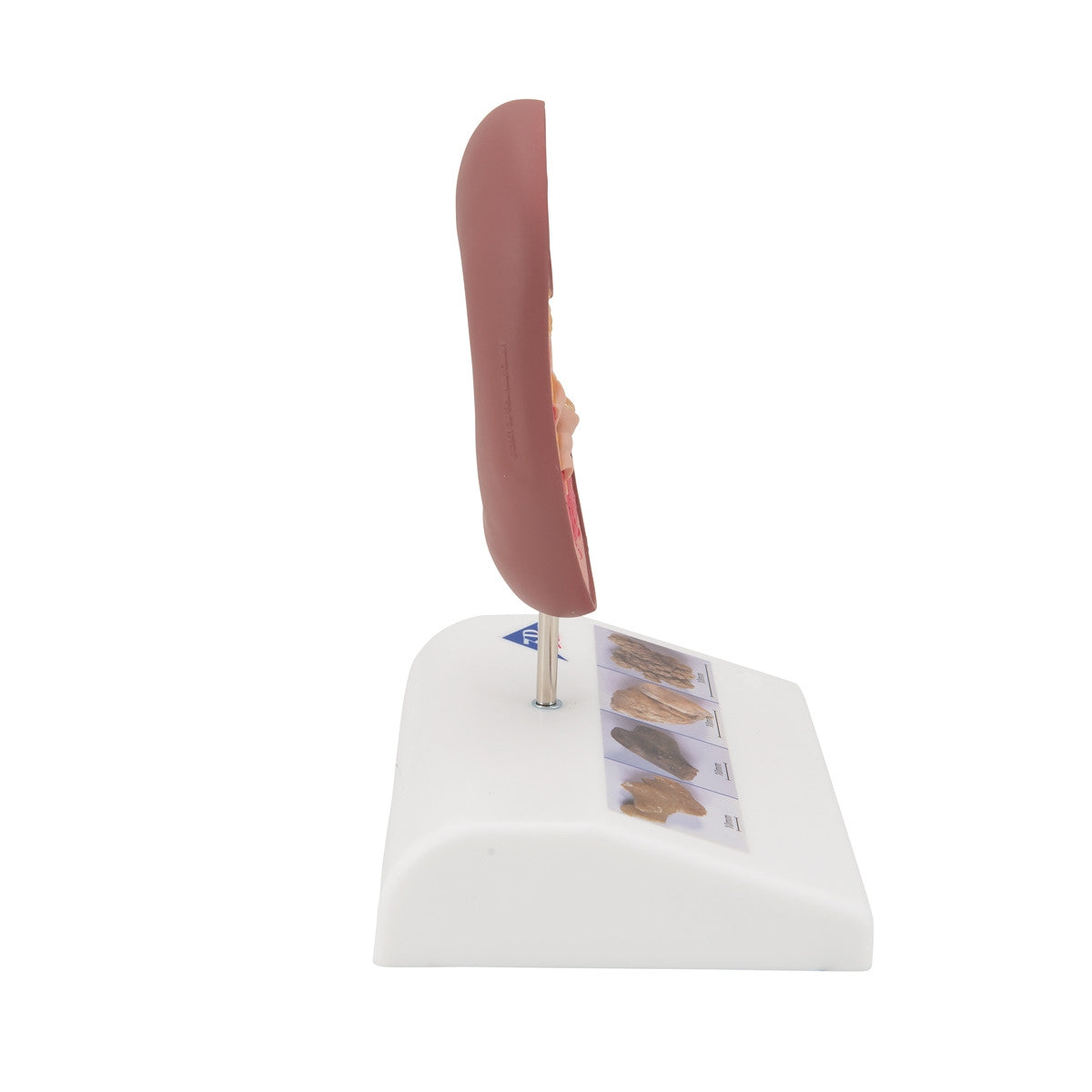
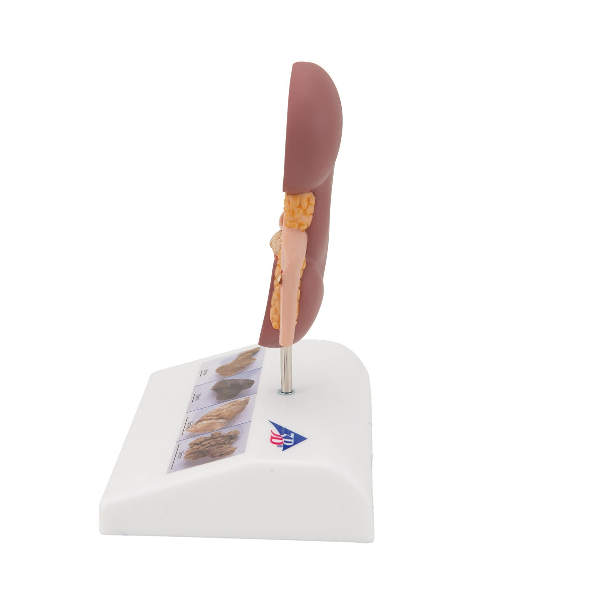
This Kidney Stone Anatomical Model comes mounted on a base with 4 illustrations of various kidney stones.
Download Kidney Stone Anatomical Model K29 / 1000316 product manual here.
This model comes with 3B Scientific 3B Smart Anatomy app included for FREE. This features access to an anatomy course, including 3 digital anatomy lectures, 117 different virtual anatomy models and 39 anatomy quizzes. It also offers a FREE warranty upgrade from 3 to 5 years with every product registration. To unlock these benefits, scan the label located on your 3B Scientific anatomy model and register online.