


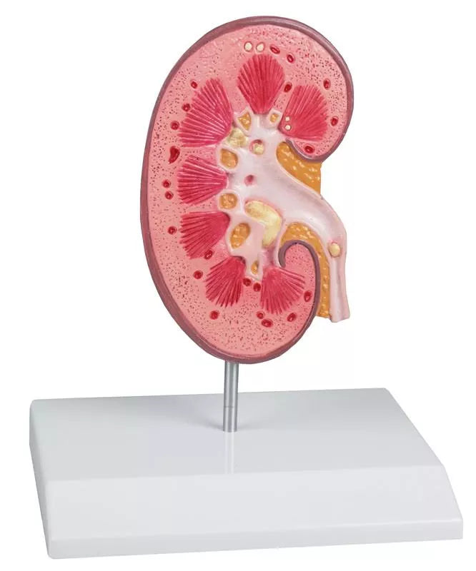
This Kidney Stone Model provides a detailed, life-size representation of a right kidney, specifically designed to support patient education on urinary stones (urolithiasis) and kidney stones (nephrolithiasis). The model is sectioned and opened to clearly display the internal anatomical structures, offering a realistic and easy-to-understand visualization of stone formation and obstruction.
The renal pelvis, renal calices, and ureter are opened to reveal concretions and stones in several typical clinical locations. These include the origin of the upper calyx group, the connecting tubule of the lower calyx group with resulting congestion of the minor calices, the renal cortex, the renal pyramids, and the ureter. Each area is carefully detailed to illustrate how and where stones commonly develop and become lodged.
Mounted on a sturdy base and supplied with a key card for easy identification of structures, this model is an excellent educational tool for medical practices, clinics, and teaching environments, helping to clearly explain the causes, locations, and effects of kidney stones.
Augmented Anatomy App Included
Enhance your learning experience with the innovative Augmented Anatomy App, designed to work seamlessly with this high-quality model. Simply point your device at the model, and the app automatically recognizes it, displaying anatomical labels and terminology in real-time augmented reality.
App Features
- High-quality augmented reality learning tool
- Free to use, with no registration required
- Anatomical nomenclature available anytime and anywhere
- Additional online resources included in the learning lexicon
- Compatible with all major smartphones and tablets
This model is ideal for anatomical study and display in GP clinics, hospitals and universities.