


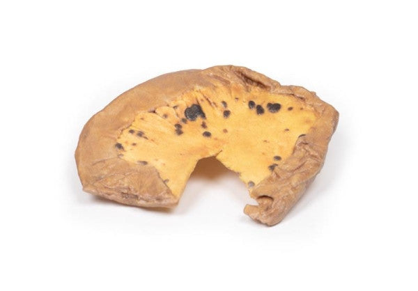
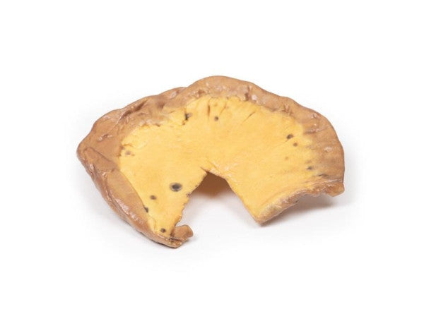
Mesenteric Metastases from Cutaneous Malignant Melanoma
The specimen consists of a segment of the small intestine arranged to exhibit the mesentery, housing multiple circumscribed nodules ranging in size from a pinhead to about 1 cm in diameter. Histological analysis verified the presence of metastatic melanoma.
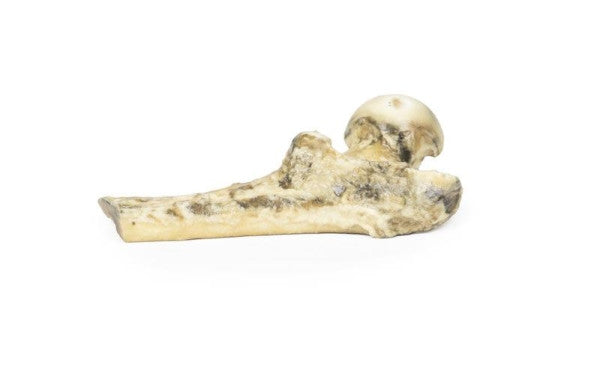
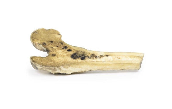
Metastatic Malignant Melanoma
The specimen is the upper right part of a right thigh bone that has been cut lengthwise to reveal tumour deposits in the marrow, ranging from light brown to black, with larger deposits destroying cancellous bone. Diffuse pale brown tumour infiltration is present, sparing the cortical bone except for medial discolouration and thickening at the shaft-neck junction. These are metastatic deposits from a skin melanoma.
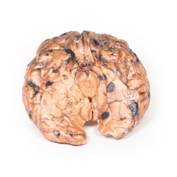
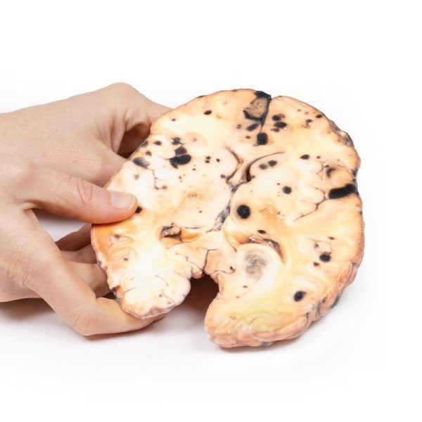
Metastatic Melanoma
This specimen showcases widespread melanoma metastases in the brain, featuring dark nodules up to 1.5 cm on both inferior and superior surfaces. The tumours invade the grey matter without encapsulation, leading to necrosis and haemorrhage.