The Osteochondroma 3D Printed Anatomy Model specimen consists of the distal portion of the patient's right femur, sectioned coronally and presented to exhibit its outer surfaces. An elongated bony growth, measuring 2 cm in length, extends from the medial side of the femoral shaft, positioned 7 cm above the medial condyle. This projection comprises typical bone tissue with a slender covering of hyaline cartilage at its apex, indicative of an osteochondroma.


Exclusive


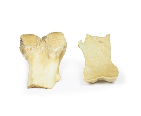
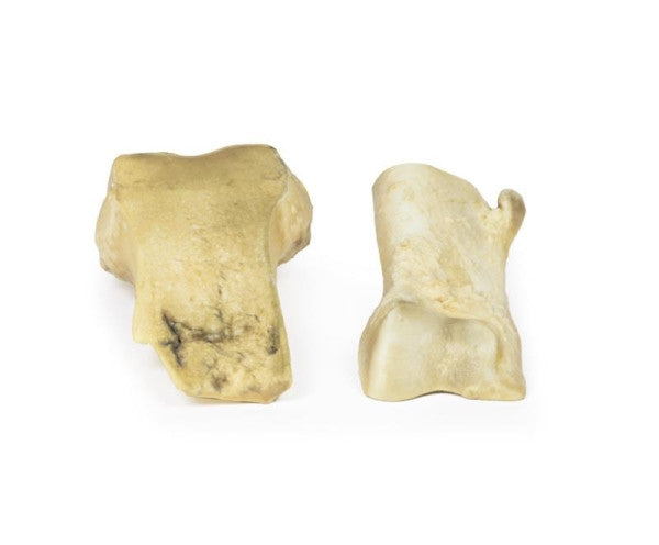
Osteochondroma 3D Printed Anatomy Model
Brand: Erler Zimmer | Made in Germany
We Accept Purchase Orders
Send your purchase order directly to us. Email orders@anatomystuff.co.uk
Price Match Service
Should you find the same item at a lower price elsewhere, please contact us.
Warranty Information
This product comes with a 3 Year warranty
Technical Information
Delivery
Returns Policy
Please note: The products featured on our website are designed solely for medical training and educational purposes.