


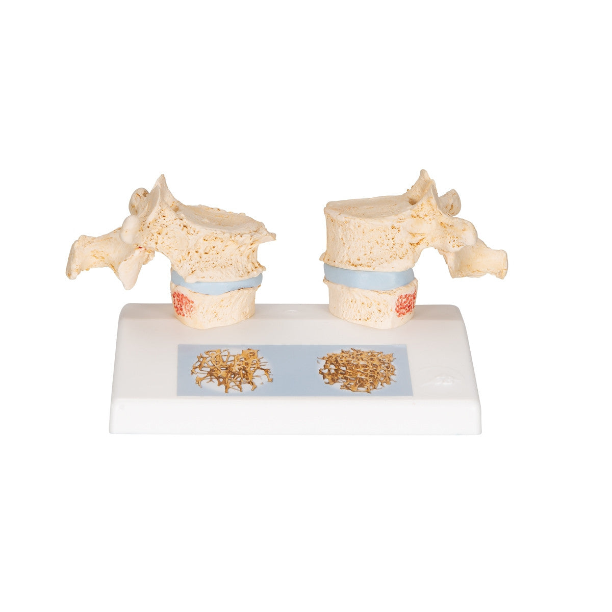
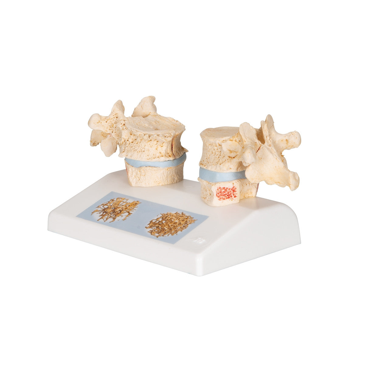
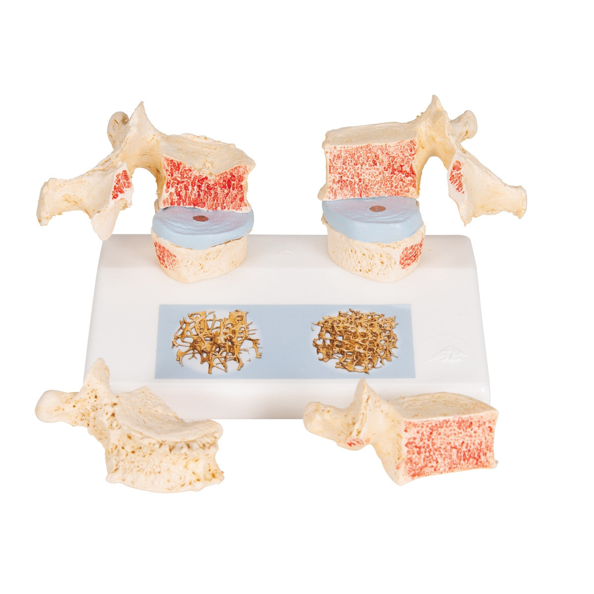
The Osteoporosis Spine Model is an accurate replica designed for comparing osteoporotic and normal thoracic vertebrae. Model A95 / 1000182 features the 11th and 12th thoracic vertebrae.
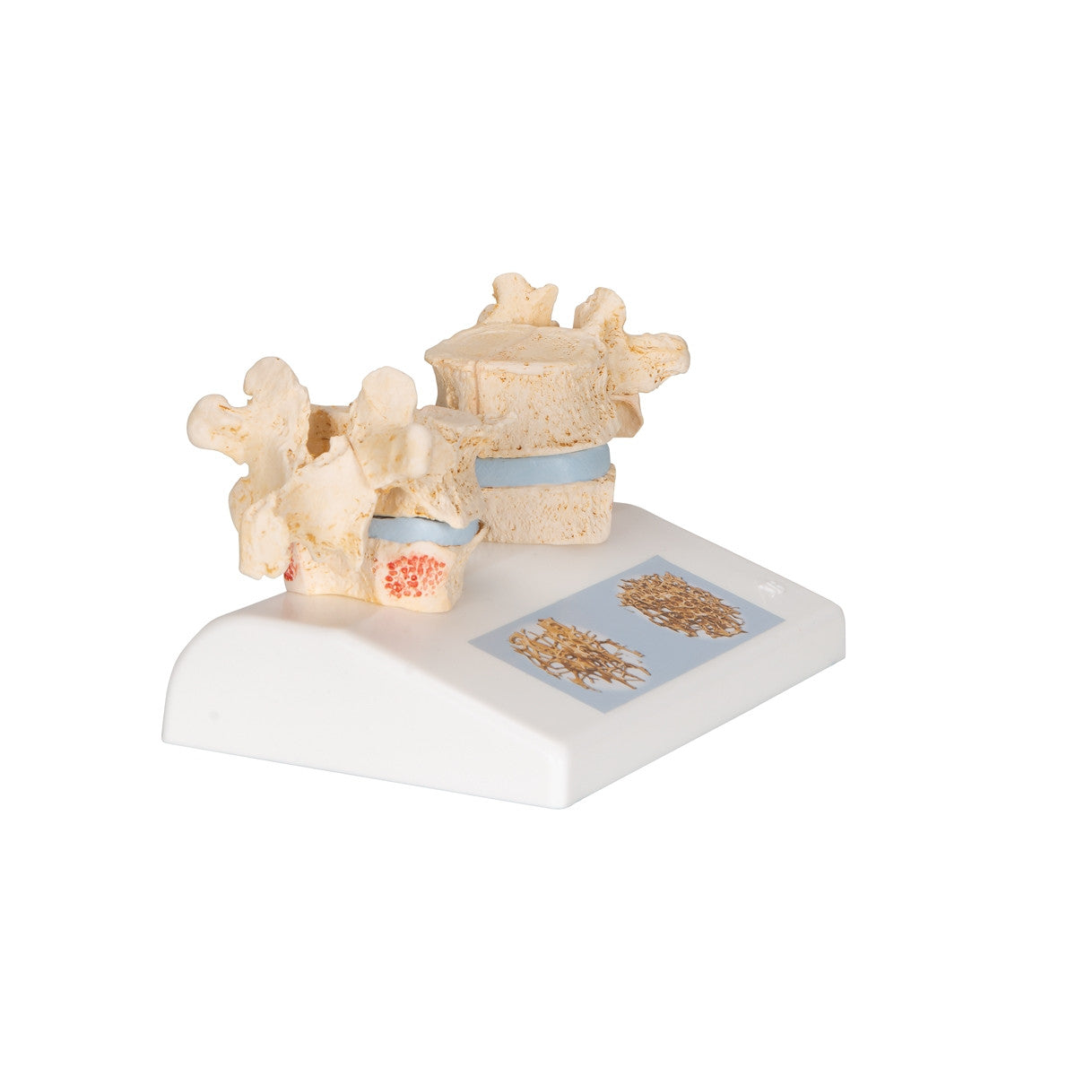
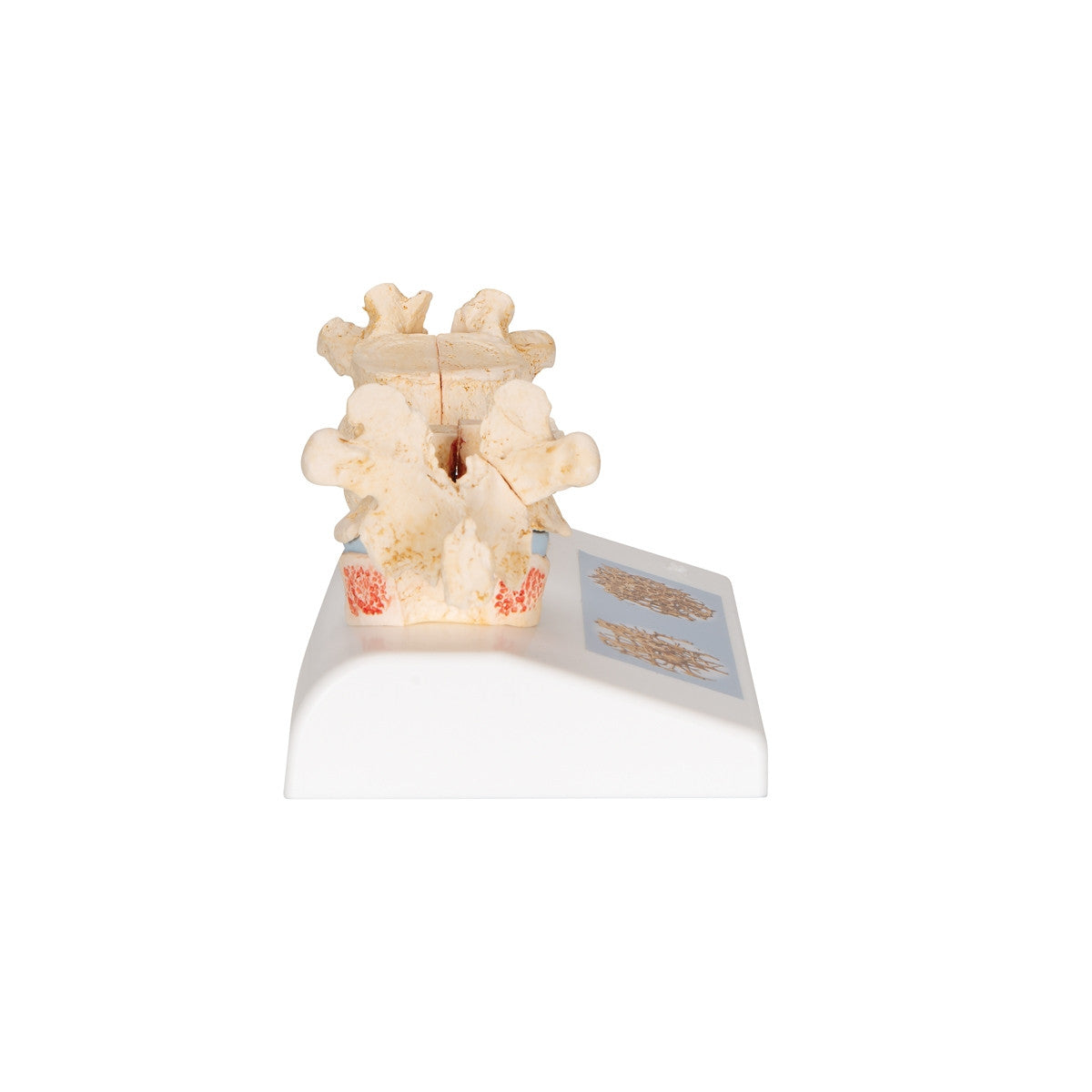
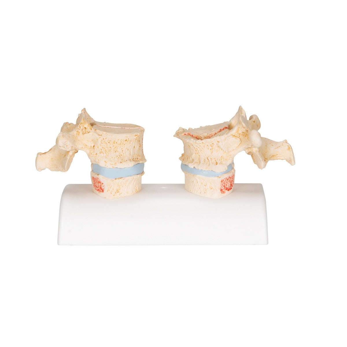
On the left of the stand reproductions of sequential osteoporotic thoracic vertebrae with narrower intervertebral discs are featured. The upper vertebrae has been split in the middle.
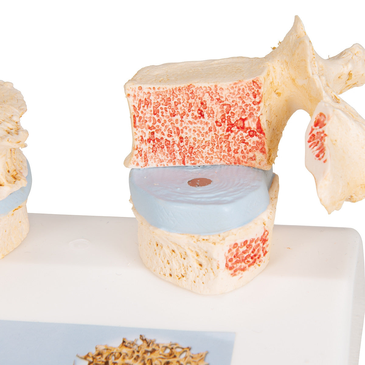
To allow for study of the fractured upper part of the vertebral body caused by sintering, such as collapse of the bony substance, the magnetically attached vertebral half can be simply detached, showing the cut surfaces. Osteophytes and other degenerative changes to the bone are also visible.
On the right hand side of the stand are two healthy vertebrae with intervertebral disc which allows for comparison. This side also features a removable half on the upper vertebral body.
On the base is a reproduction of two 3D micro CT images taken from bone biopsies, illustrating the microarchitecture of an osteoporotic bone and showing difference in bone density in relation to a healthy bone.
A very detailed osteoporosis anatomy model.
Download Osteoporosis of the Thoracic Spine Model A95 / 1000182 product manual here.
This model comes with 3B Scientific 3B Smart Anatomy app included for FREE. This features access to an anatomy course, including 3 digital anatomy lectures, 117 different virtual anatomy models and 39 anatomy quizzes. It also offers a FREE warranty upgrade from 3 to 5 years with every product registration. To unlock these benefits, scan the label located on your 3B Scientific anatomy model and register online.