


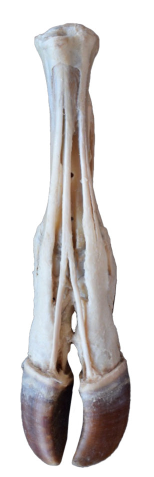
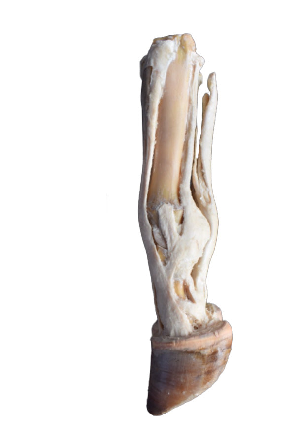
This 3D Printed Ox Foot Model illustrates the anatomy of an ox's right foot, from the metatarsus to the distal phalanges.
The plantar aspect reveals the relationship between the tendons of the superficial and deep digital flexor muscles and deep structures such as the interosseous ligament.
On the dorsal aspect, the primary structures include the insertions of the common, lateral, and medial digital extensor muscles. Additionally, the capsular and extra-capsular ligaments of the metatarsophalangeal and interphalangeal joints are preserved.
This model is part of a range of detailed 3D printed veterinary models, exclusively available in the UK via AnatomyStuff, ideal for advanced anatomical study.