


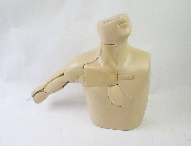

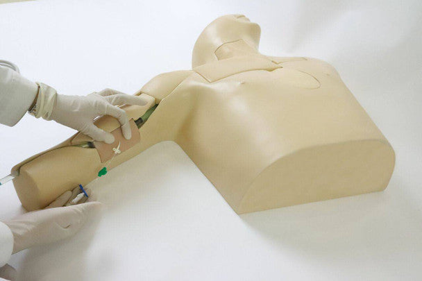


The Peripherally Inserted Central Catheter (PICC) Ultrasound Training Model has been carefully designed for the training and practice in the correct placement of a Peripherally Inserted Central Catheter (PICC) line. The model is beneficial for the use of medical students and trainee doctors.
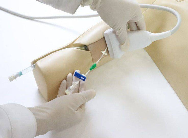
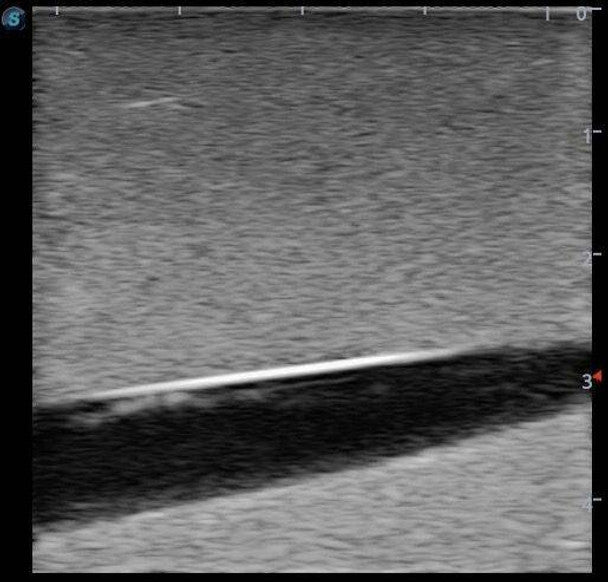
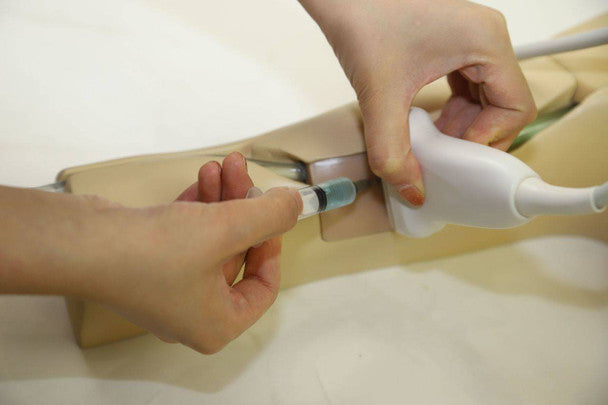
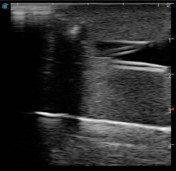
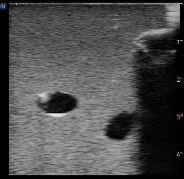
The anatomical structures that can be observed and are correctly positioned on the model are the clavicle, rib, sternocleidomastoid, jugular vein and basilic vein. The central venous catheters are inserted by the students in the identified basilic vein of the arm or the jugular vein which can be found in the neck region. A guide wire can be followed whilst inserting the catheter into the central vein and observed with an ultrasound to ensure proper placement of the catheter.
The model is made of a high molecular polymer ultrasound material and is a close representation of real skin, therefore actual ultrasound technology can be used. The modules of the puncture positions are replaceable, meaning procedures can be repeated and the model can be reused by students.
Essential skills that are gained from this training model are:
- Peripherally inserted central catheter (PICC) training
- Trocar puncture
- Skin dilation using an expander
- The correct use of an ultrasound and the interpretation of the corresponding images
- Sterile techniques
This PICC Ultrasound Training Model is realistic and an invaluable asset to clinical skills sessions.
Contents Included
- 1 x PICC ultrasound training model
- 1 x 150ml Red contrast solution
- 1 x 150ml Blue contrast solution
- 1 x 50ml Syringe
- 1 x Water inlet connector
- 1 x Operation manual