


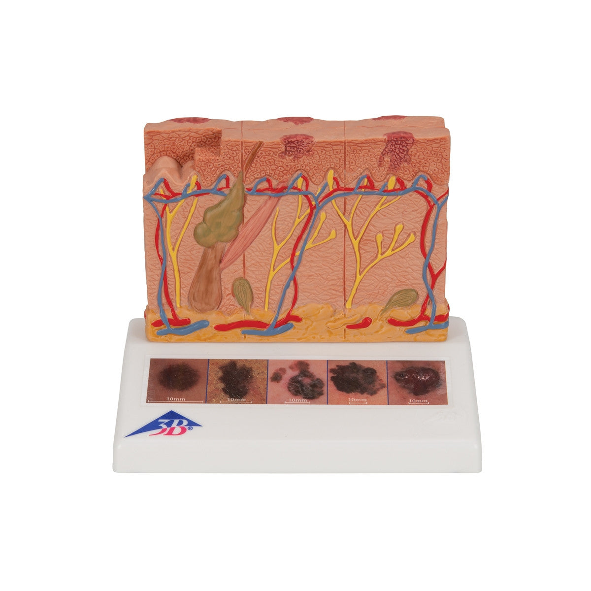
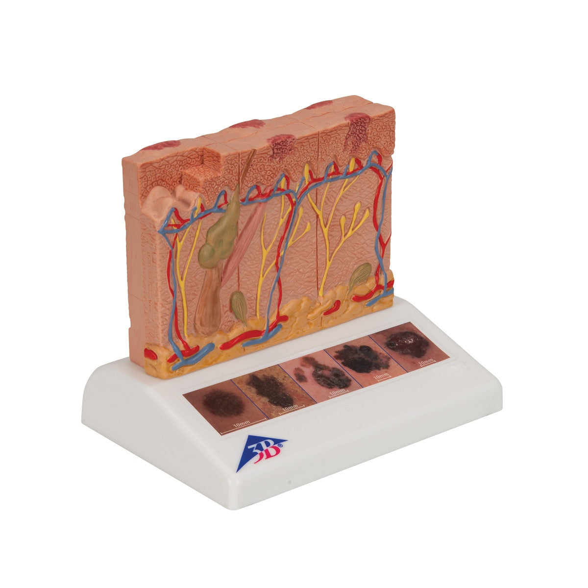
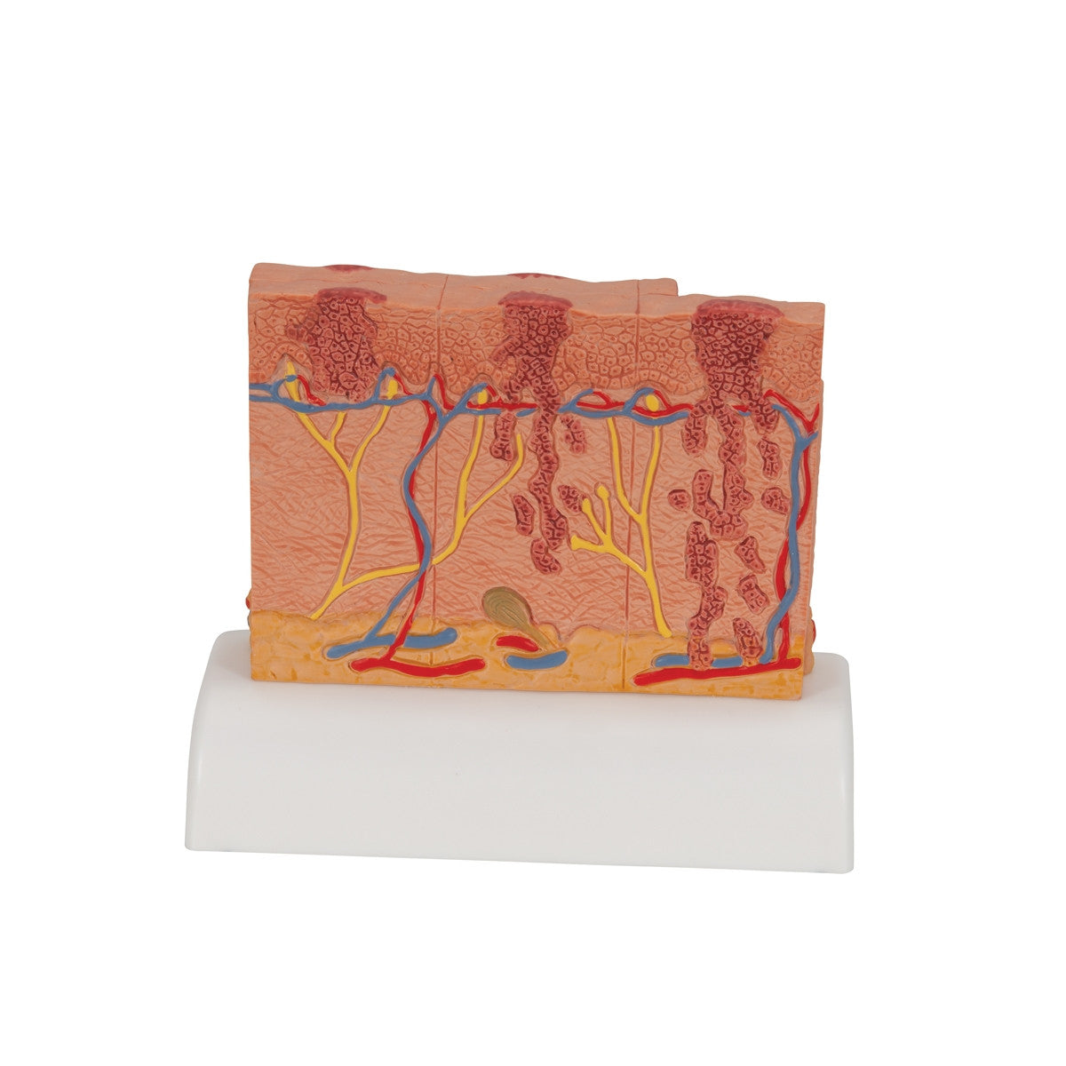

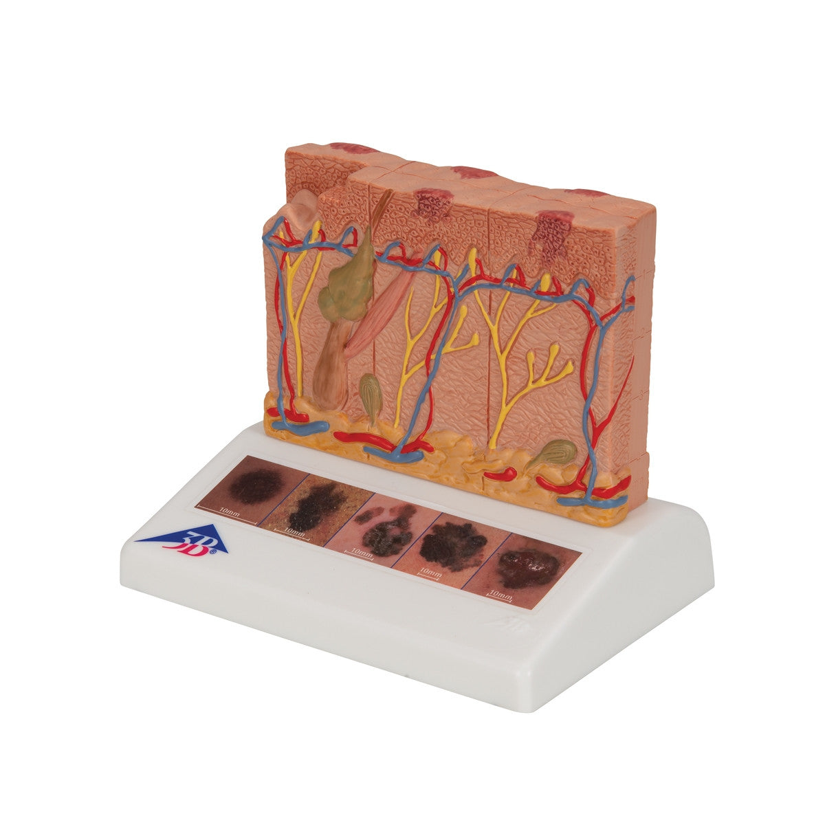
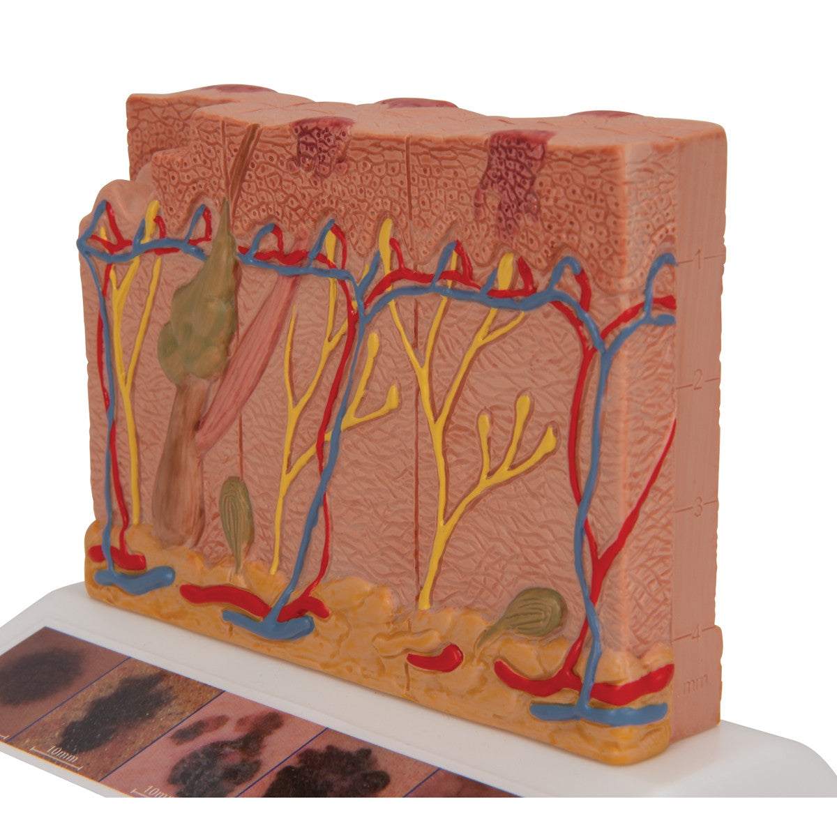
Enlarged skin model showing both healthy skin and five different stages of malignant melanoma on the front and back.
This skin cancer model presents both healthy skin and five different stages of malignant melanoma on the front and back, which have been enlarged to 8 times life-size for easier study. The stages presented by model J15 / 1000293 are as follows:
- healthy
- malignant cells are found at the surface, within the epidermis
- malignant cells fill the epidermis, a few invade the papillary layer
- malignant cells fill the papillary layer
- malignant cells invade the reticular layer
- malignant cells have reached the subcutaneous fatty tissue, satellite cells approach a vein
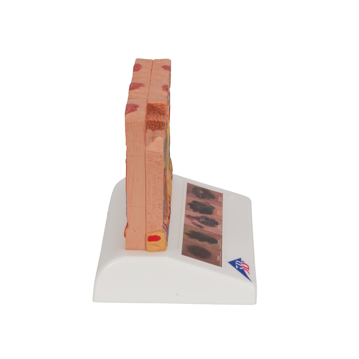
The top view of this skin cancer model reveals external visible skin changes, allowing for an assessment according to the "ABCDE" criteria. While the side of the model features the different levels of invasion into the skin layers according to Clark (I-V) and the tumour thickness in mm according to Breslow. Included on the base are five colour illustrations of types of malignant melanomas making this model an ideal skin pathology educational tool.
Download Skin Cancer Model (8 times life size) J15 / 1000293 product manual here.
This model comes with 3B Scientific 3B Smart Anatomy app included for FREE. This features access to an anatomy course, including 3 digital anatomy lectures, 117 different virtual anatomy models and 39 anatomy quizzes. It also offers a FREE warranty upgrade from 3 to 5 years with every product registration. To unlock these benefits, scan the label located on your 3B Scientific anatomy model and register online.