


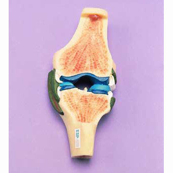
This rigid life-size model shows the anatomy of a synovial joint in detail. The bone structure is displayed in cross-section so that the articular disc, articular cartilage, synovial membrane and capsular ligament are visible.
Parts of the synovial joint are colour-coded to assist identification and certain sections have been removed to reveal the underlying structures.