


Discover this hands-on and highly realistic Thoracentesis Ultrasound Training Model. It is an excellent resource for healthcare students and professionals.
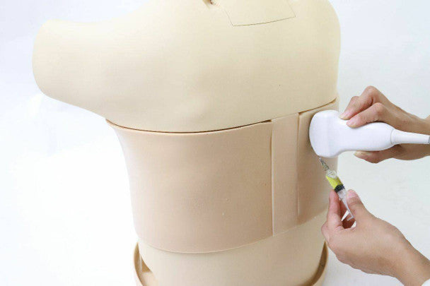
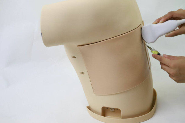
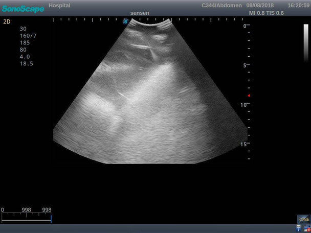
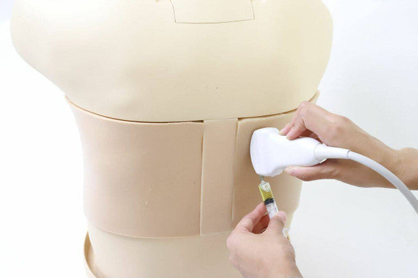
Trainees can first examine the patient, palpating key anatomical landmarks. They can also use the ultrasound probe to recognise landmarks by image interpretation. These images are real clinical ultrasound images with clear muscles, connective tissues, lungs and pleural structures.
Next, students can perform an important clinical skill: an ultrasound-guided thoracentesis. They can insert the needle and aspirate fluid if the puncture is successful. They will also feel resistance and ‘pop’ through the needles. This makes the learning experience extremely realistic.
Due to the generous reservoir (300ml) in both sides of the models, trainers do not have to worry about refilling the fluid often, guaranteeing prolonged and extended sessions. Additionally, the puncture pad is durable and replaceable for repeated use.
The model is compatible with some real ultrasound machines.