


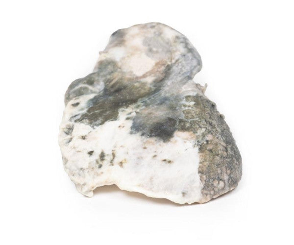
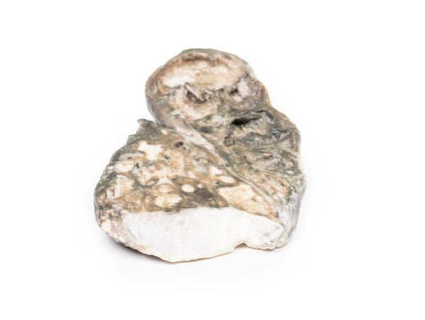
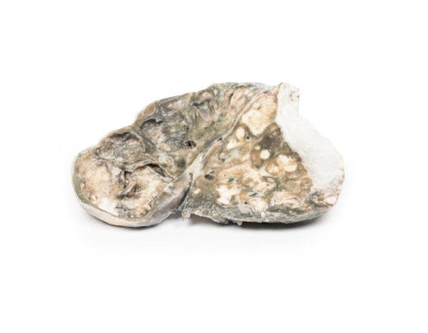
Fibrocaseous Tuberculosis
The left lung is longitudinally cut, displaying a replaced upper lobe with irregular cavities containing necrotic debris. Haemorrhage is evident in the upper cavity, while the lower lobe shows smaller caseous areas breaking down, with scarred lung tissue. Thickened pleura indicates fibrocaseous tuberculosis with cavitation.
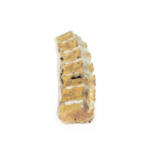
Tuberculosis
The specimen shows a sawn portion of a thoracic vertebral column, displaying the cut surface of seven thoracic vertebrae, with osteolytic areas containing mostly lost caseous degenerative material, surrounded by a thin zone of dense bone. Tuberculosis inflammation has extended into an intervertebral disc and formed collections of caseous material beneath the anterior longitudinal ligament. This represents an instance of tuberculous mycobacterial osteomyelitis (Pott's Disease).