


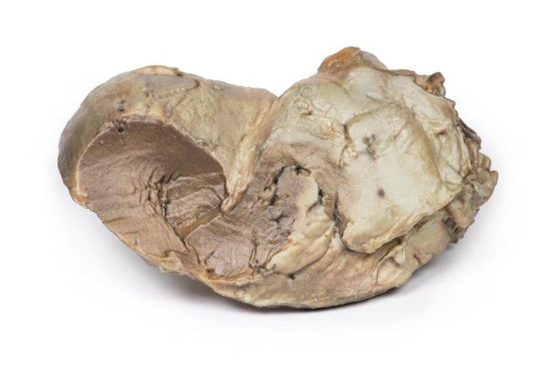
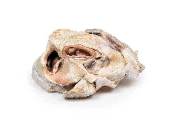
Bicuspid Aortic Valve
The heart has been opened to reveal the left ventricle and associated valves. The aortic valve has two cusps instead of the typical three, with slight patch thickening. The left and right coronary arteries' aortic origins are widely open, along with the left circumflex coronary artery. The specimen exhibits dense pericardial fibrosis and adhesions on the posterior side, indicative of constrictive pericarditis. The cause is unclear from the provided history.
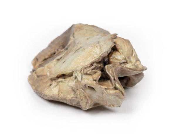
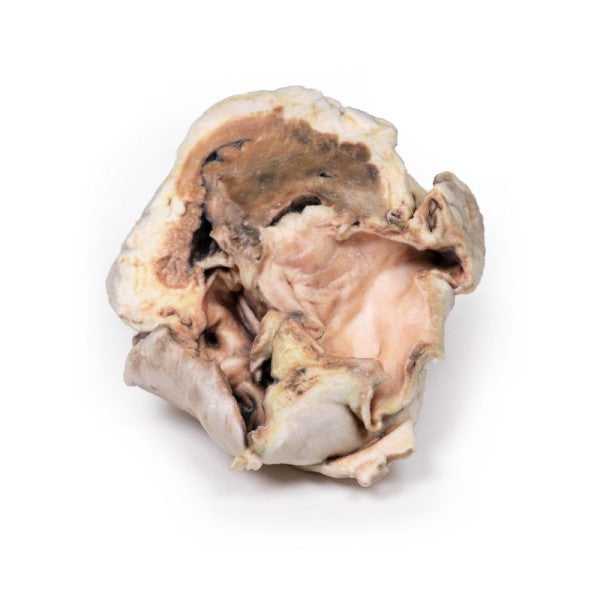
Calcified Aortic Valvular Stenosis (Bicuspid Aortic Valve)
The specimen is a cross-section of the left atrium, revealing the smooth internal lining, left auricular appendage, and part of the left ventricle on the lower aspect. On the upper aspect, the pulmonary trunk, part of the pulmonary tricuspid valve, and the aorta with an abnormal bicuspid valve are visible. Calcified aggregations on the valve margins and a region of calcification on one cusp of the pulmonary valves are evident from this perspective.
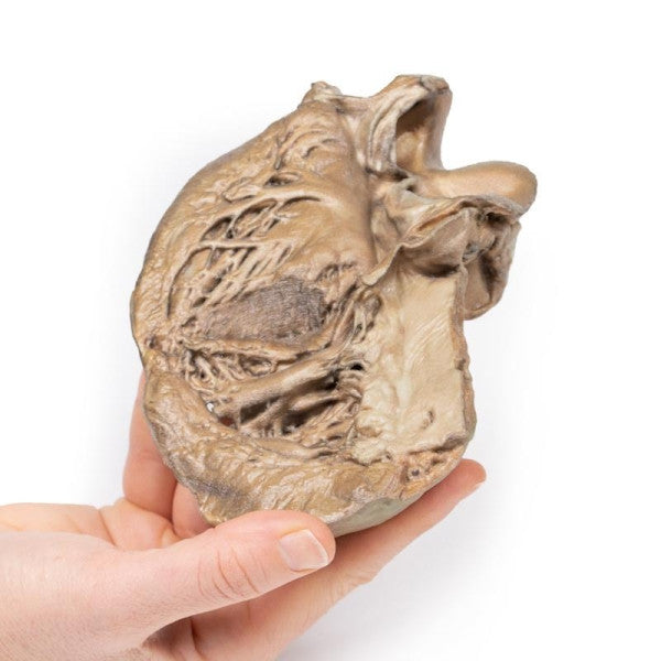
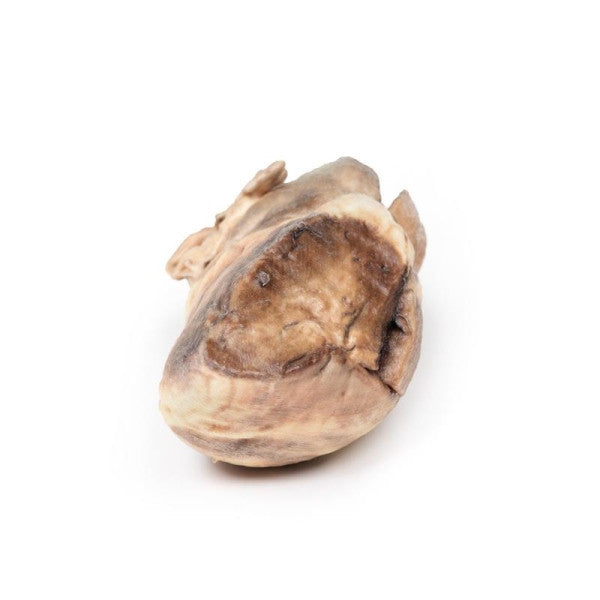
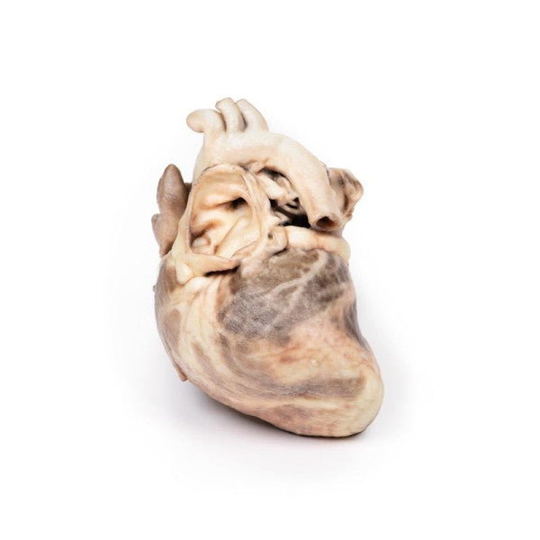
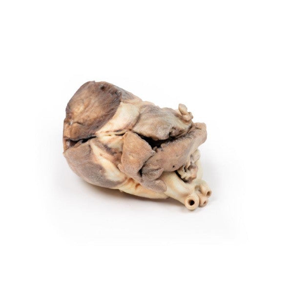
Congenital Pulmonary Stenosis
The child's heart specimen shows an abnormal pulmonary valve with a thickened conical diaphragm and a 2mm opening at the apex. The pulmonary artery displays significant post-stenotic dilatation. Right-sided cardiac enlargement is observed due to marked dilatation of the right atrium and auricle, along with right ventricular hypertrophy, visible through incisions in the myocardium. The diagnosis is pure pulmonary valve stenosis.