


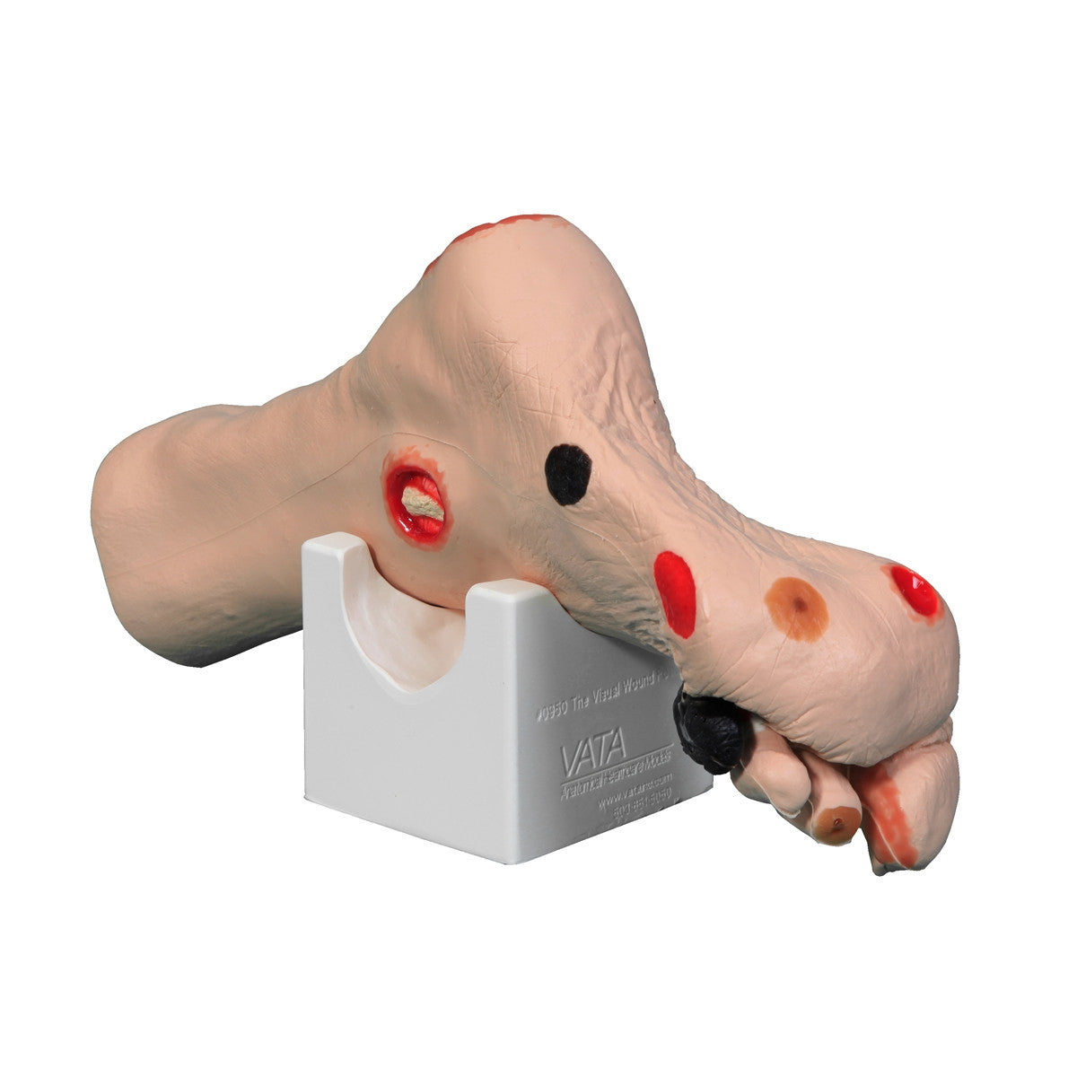
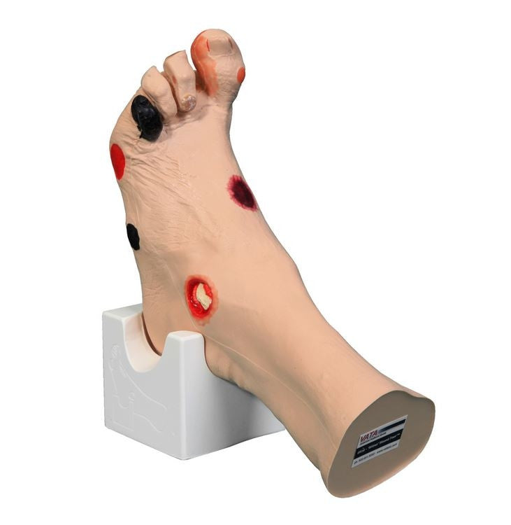
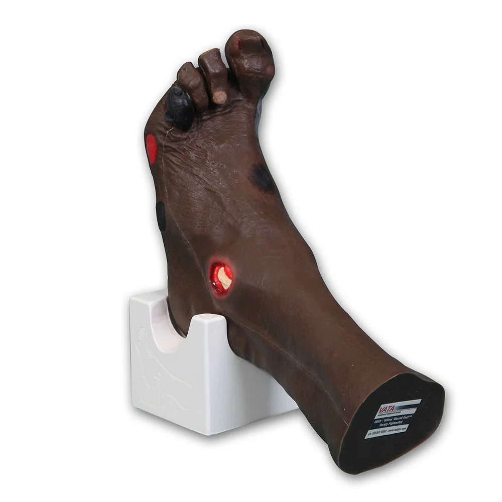
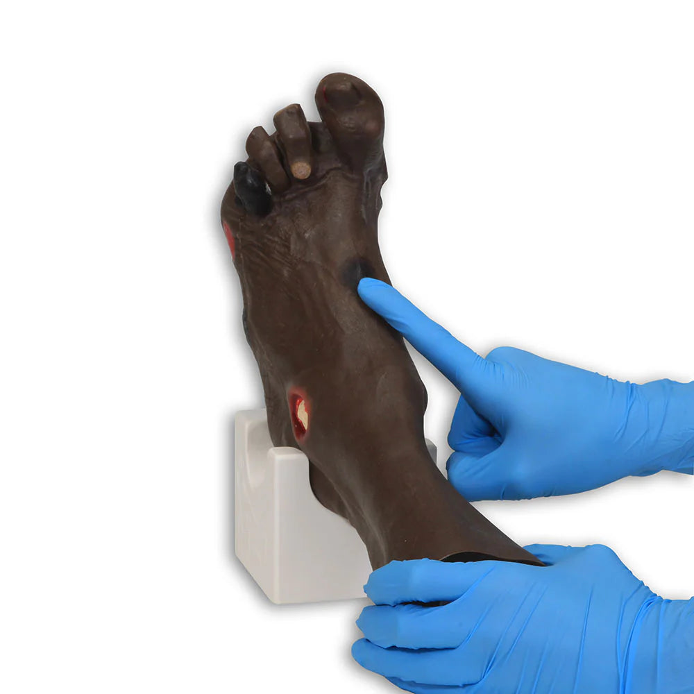
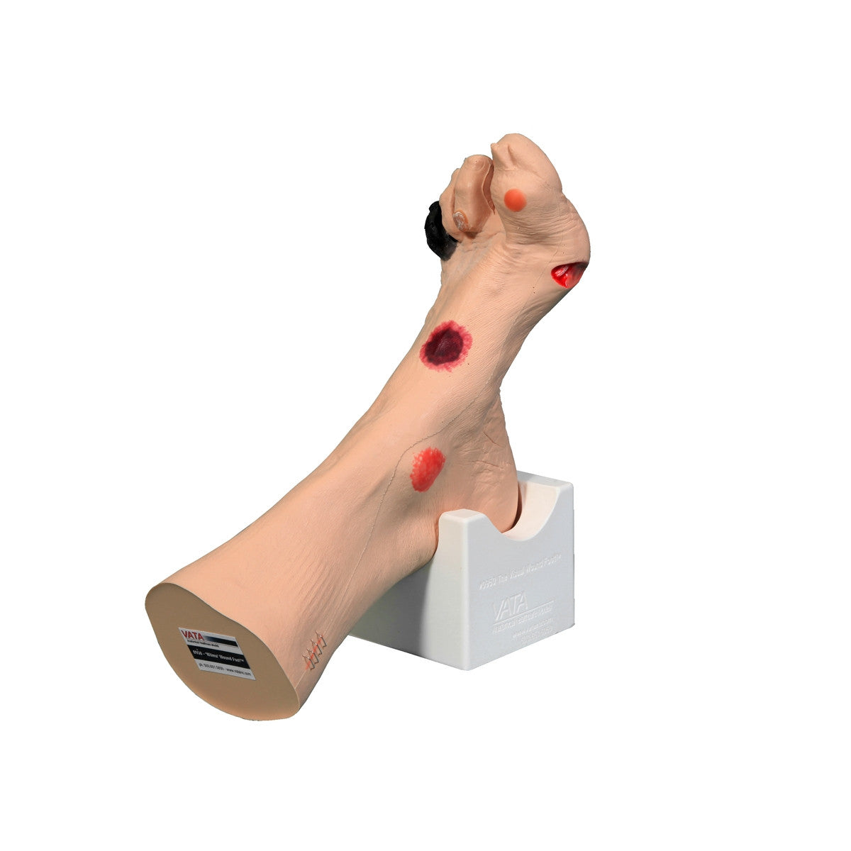
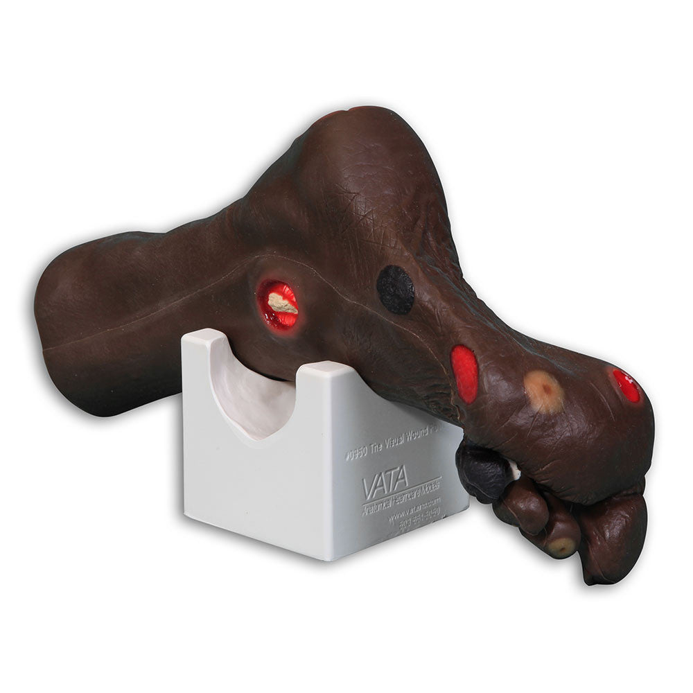
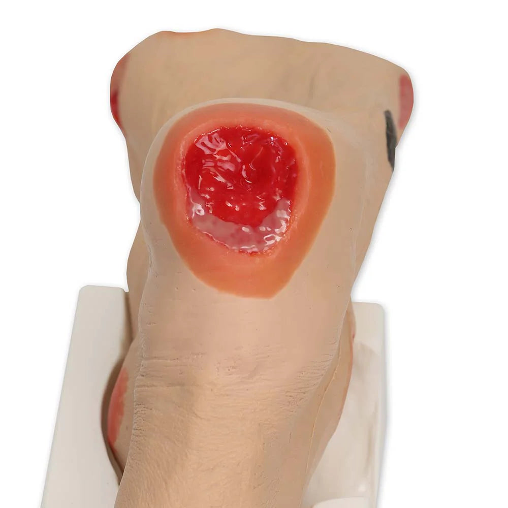
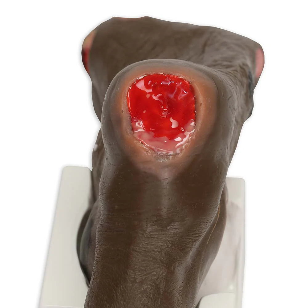
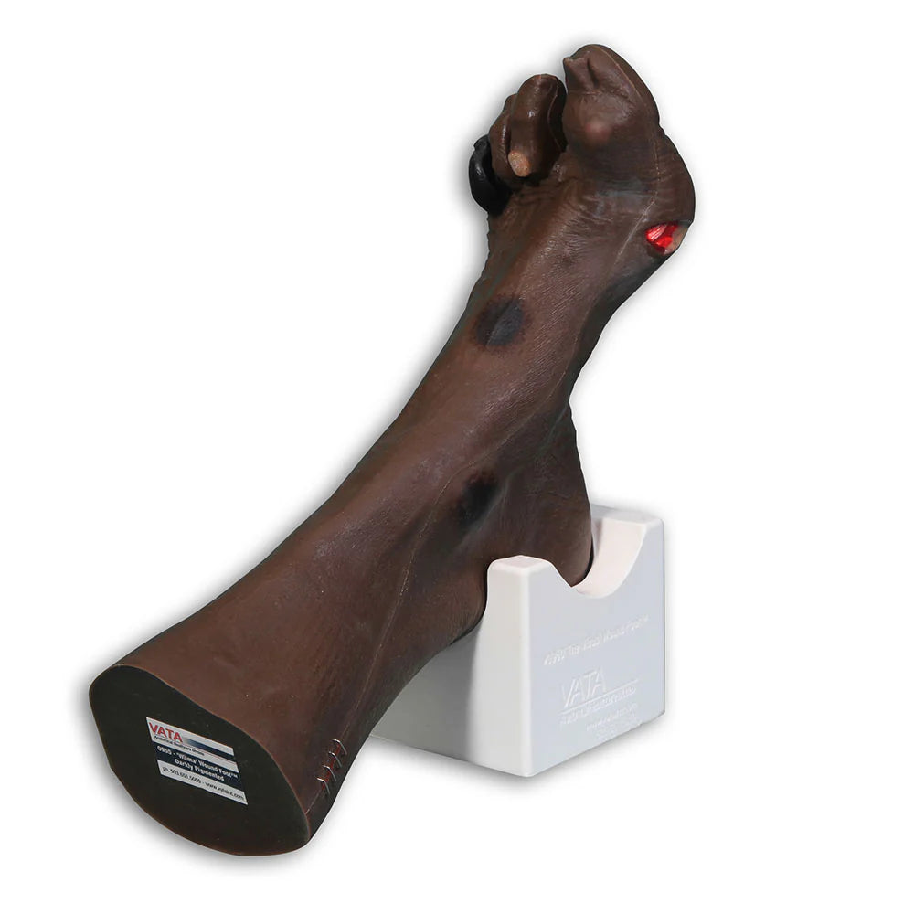
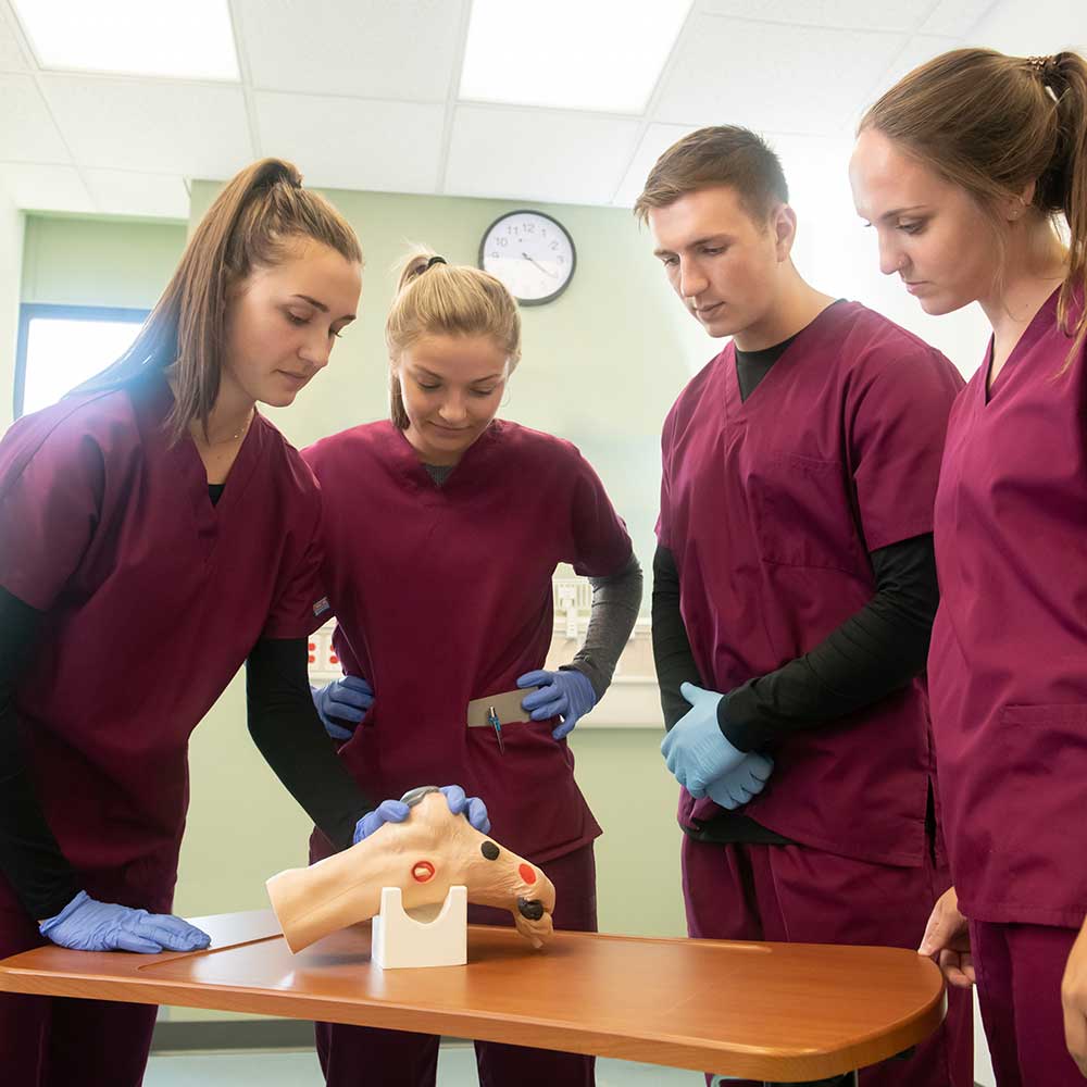
A comprehensive visual wound foot model based on a mould from an 80-year old patient. The clinical skills trainer by VATA offers health professionals a true-to-life experience of assessing the various wounds. Twenty different conditions are presented on Wilma Wound Foot with accurate colouration, to help students identify the probable etiology of different wounds and devise effective treatment plans.
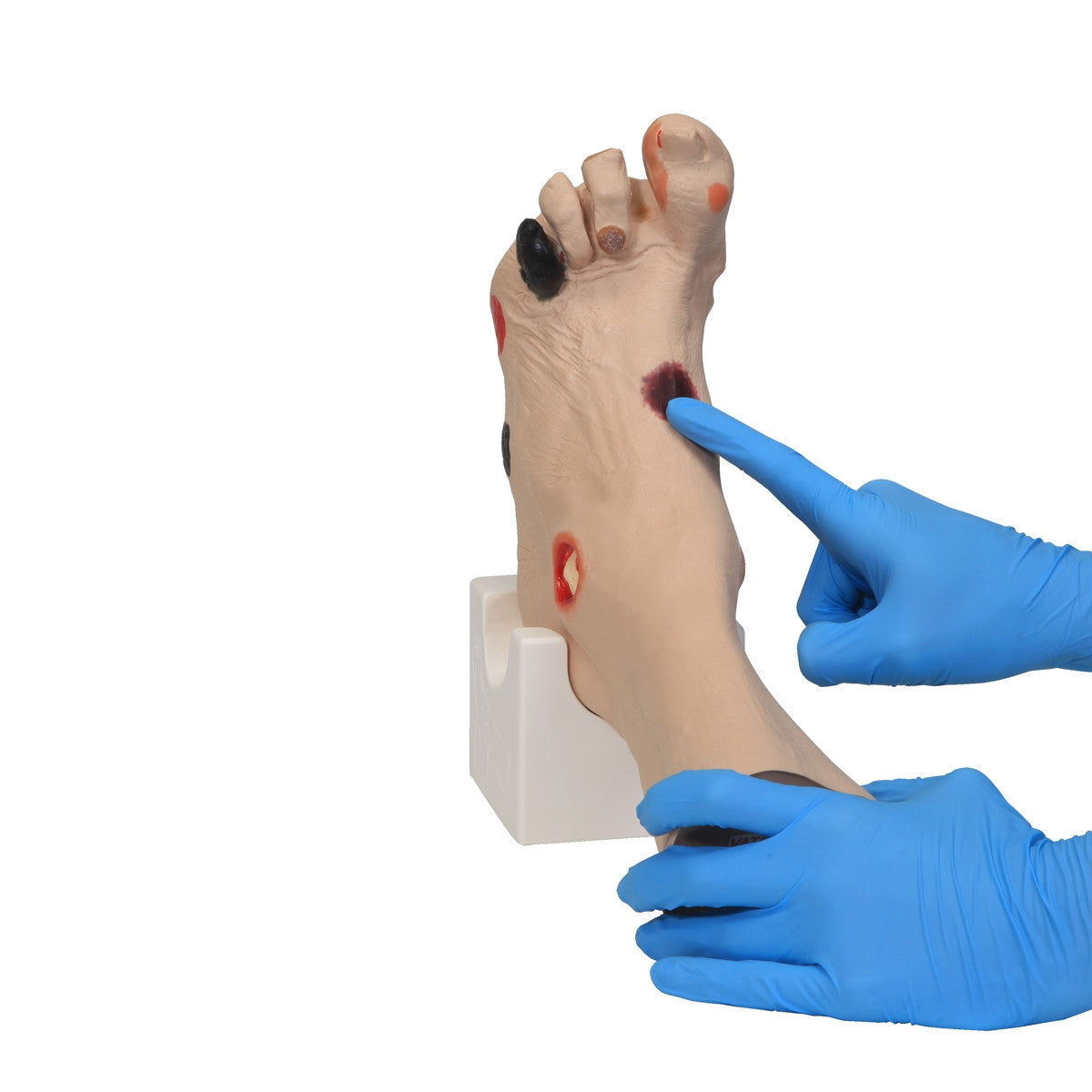
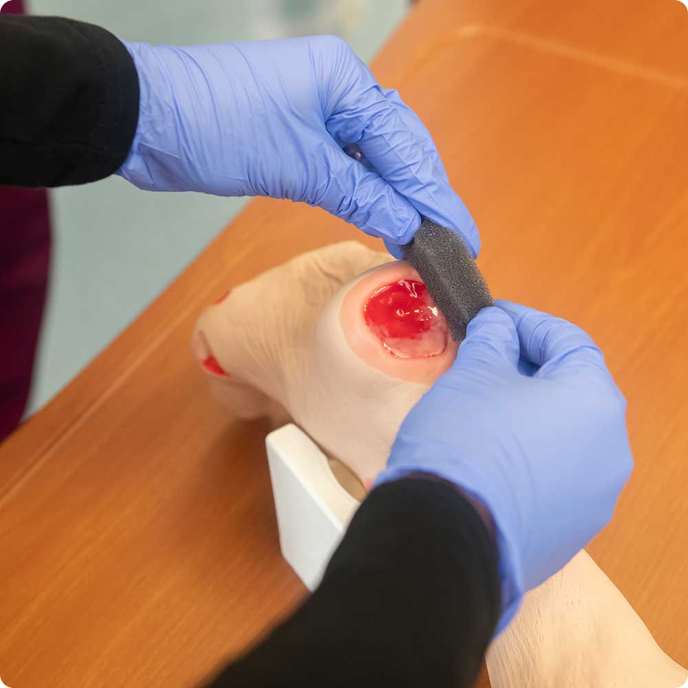
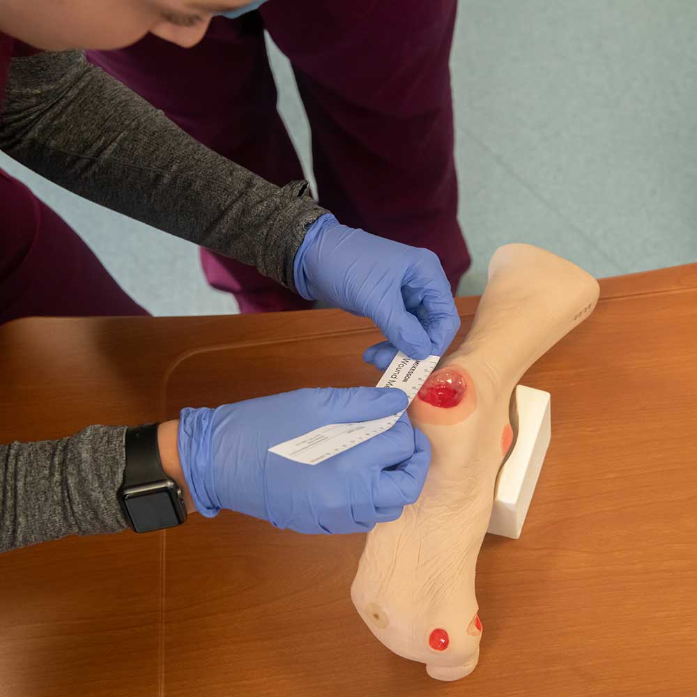
The Wilma Wound foot can also help patients in the identification and staging of wounds and their probable etiologies. Wilma Wound Foot is also an excellent visual aid for educating those who cannot read well enough to understand basic health care information, allowing them to see what can occur without proper care. Routine cleansing and dressing changes can be taught and practiced on all the wounds by healthcare providers, patients, families and caregivers.
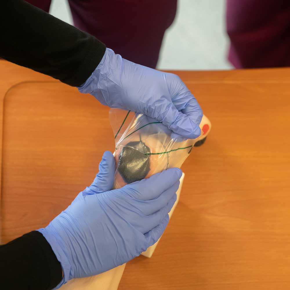
The model is made of flexible material permitting the toes to be moved for closer examination or the application of dressings, and the Wilma foot model is supplied with a positioning base which will give 'hands free' access to all the sites when applying dressings or for teaching.
The following wounds, pressure ulcers and foot abnormalities are presented on Wilma Wound Foot :
- Stage I pressure ulcer on the medial malleolus
- Stage II pressure ulcer on the lateral foot (behind the 5th toe)
- Stage III pressure ulcer on the heel with infection
- Stage IV pressure ulcer on the lateral malleolus with exposed tendon and bone (with osteomyelitis)
- Stage IV pressure ulcer on the medial foot (behind the great toe) with exposed tendon and slough
- Suspected DTI (Deep Tissue Injury) on top of foot with 'mushy/boggy' feel when palpated
- Unstageable eschar on lateral foot * Full thickness ulcer, plantar, 1st digit, probable diabetic neuropathic etiology
- Callus under the 4th metatarsal phalangeal joint
- Amputated 2nd toe * Dry gangrene of the 5th toe, due to ischemia
- Interdigital maceration between the 4th and 5th toe
- Partial thickness wounds between 3rd and 4th toe at 1st joint
- Corn on top of 3rd toe
- Callus at tip of 3rd toe
- Ingrown toenail on the medial aspect of the great toe
- Fungal thickened toenail on the great toe
- Blister on great toe at the first joint
- Hammer toes - 3rd, 4th and 5th toes
- Skin stapled wound
Please select from dark or light skin tones. An optional carry case is available to purchase.