


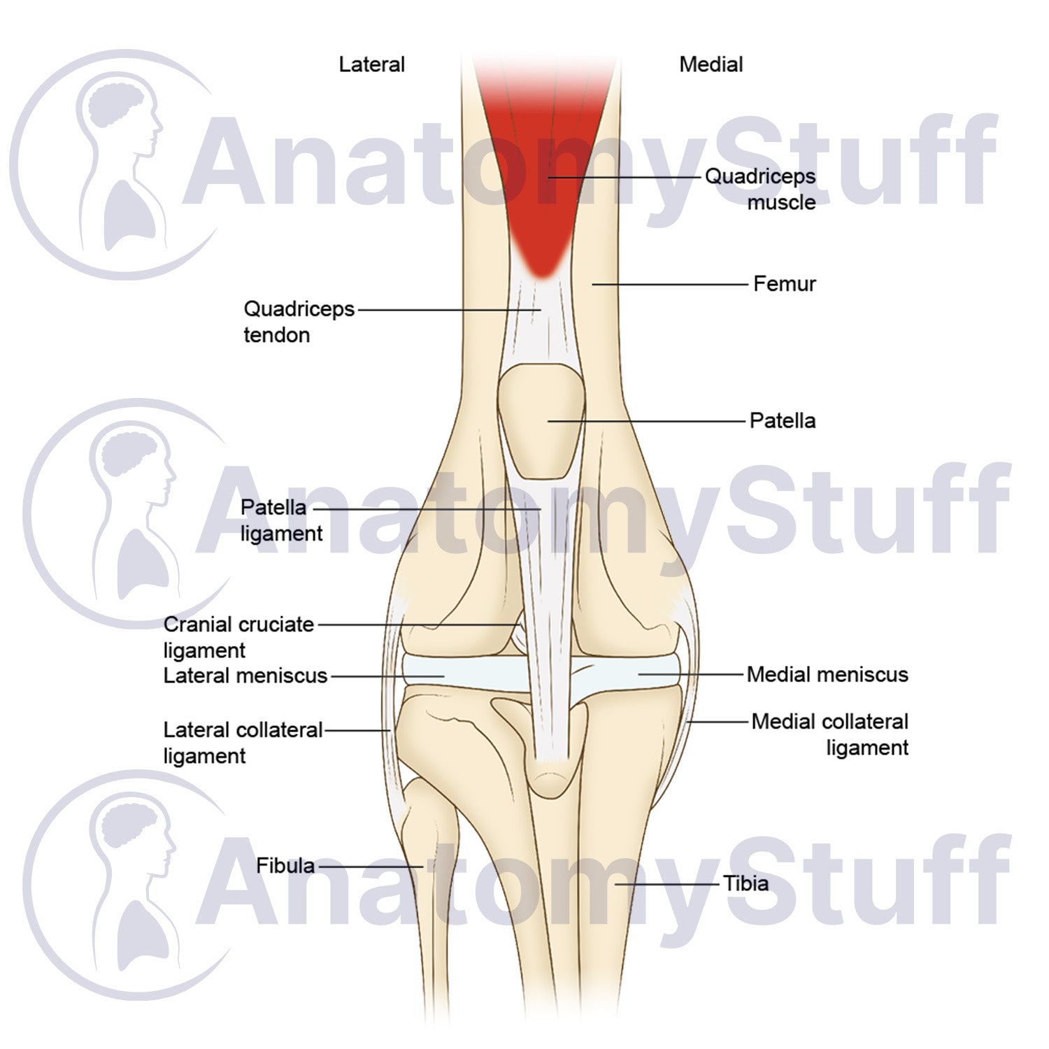
This licensable scientific illustration of Canine Stifle Joint Anatomy provides a clear, comprehensive view of the dog knee joint in a craniocaudal aspect. Illustrated by a professional medical illustrator, this stock image asset is perfect for educators, veterinary professionals, researchers, and students. Available with both labelled and blank variants for veterinary teaching, student revision, and educational presentations.
Anatomical Highlights
- Bony Structures: Detailed rendering of the femur, tibia, fibula, and patella knee cap.
- Tendons & Ligaments: Features the quadriceps tendon, patella ligament, cranial cruciate ligament CCL, lateral collateral ligament, and medial collateral ligament.
- Joint Cartilage: Accurately maps the lateral meniscus and medial meniscus structures.
- Musculature: Clear identification of the distal quadriceps muscle insert.
Choose Your Variant
- Fully Labelled: Ready for immediate use in presentations.
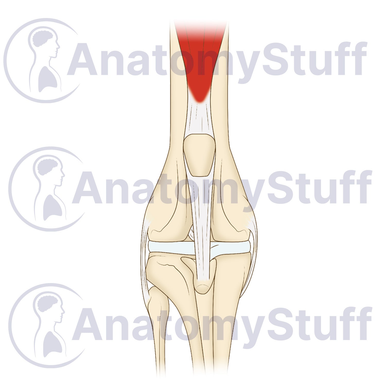
- Unlabelled (Blank): Perfect for interactive learning. This clean version is ideal for student examinations, "fill-in-the-blank" quizzes, or custom labelling for specialised research.
Product Specifications
- Format: PNG (Transparent background)
- Dimensions: 1100 x 880 px
- Resolution: 300 DPI
- Print Size: ~10 x 8 cm
- Colour Profile: RGB (Optimised for digital and print)
- File Size: ~200 KB (Blank) ~250 KB (Labelled)
Licensing Information
Please select the licence that matches your intended use:
- Science Licence: Licence for academic purposes such as theses research publishing, and the scientific discourse.
- Education Licence: Licence for educational purposes, live teaching, presentations, handouts, and exam papers.
Commercial Use: Interested in using this for advertising, book publication, or other commercial purposes? Please Contact Us to discuss a Commercial Licence.
Please allow 1-2 working days for delivery of your image.
By purchasing, you agree to our Licensing Terms and Conditions.