






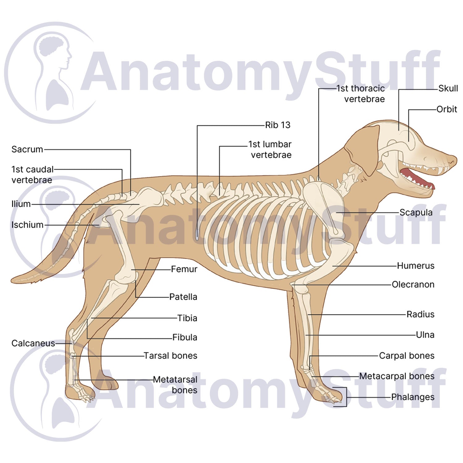
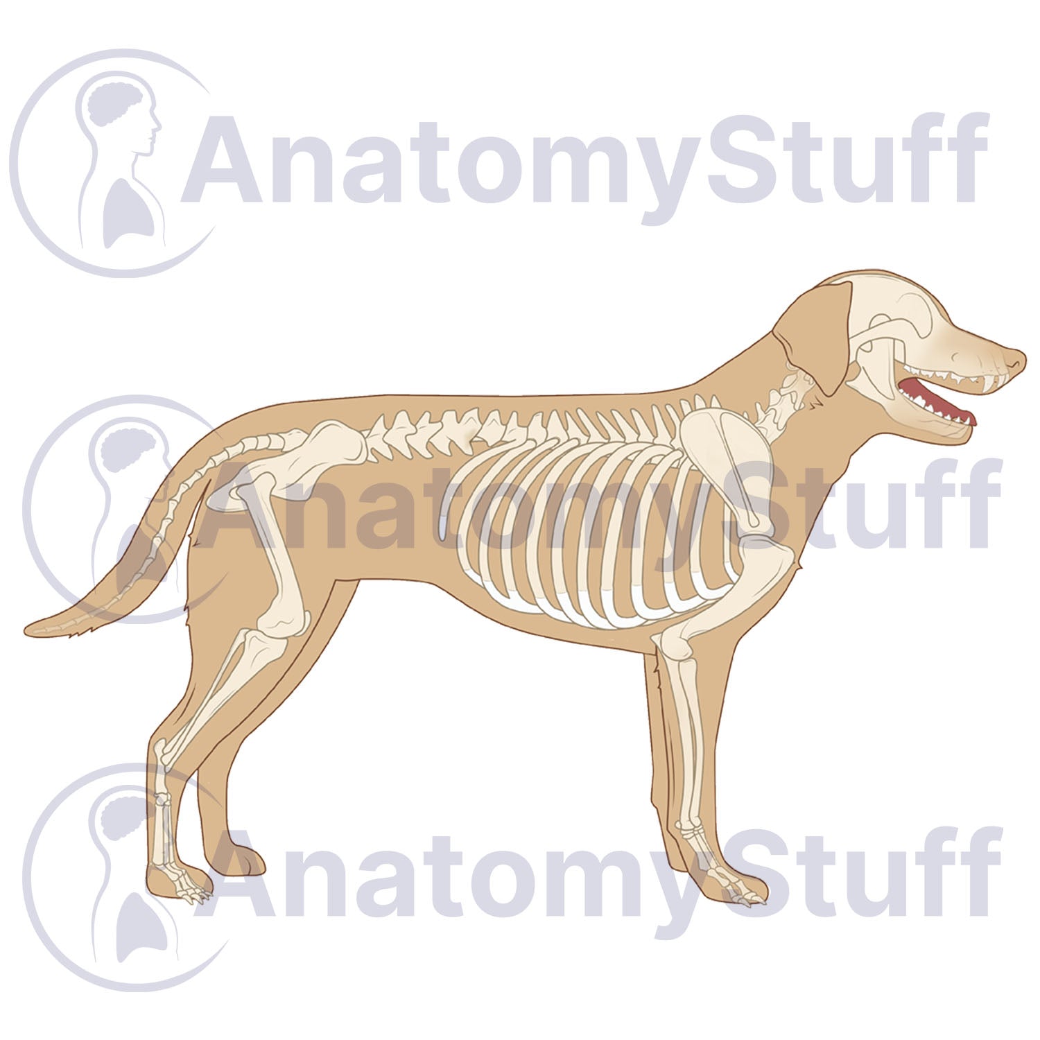


This licensable scientific illustration of Canine Skeletal Anatomy provides a clear, comprehensive view of the dog skeletal system, including the complete vertebral column and distal limb structures. Illustrated by a professional medical illustrator, this stock image asset is perfect for educators, veterinary professionals, researchers, and students.
Anatomical Highlights
- Axial Skeleton: Detailed rendering of the skull and ribs, alongside the entire spinal column including cervical, thoracic, lumbar, sacral, and coccygeal vertebrae.
- Forelimb Anatomy: Clearly illustrates the pectoral girdle and front leg bones, including the scapula, humerus, radius, ulna, carpus, metacarpals and phalanges.
- Hindlimb Anatomy: Features the pelvic girdle and hind leg bones, including the pelvis, femur, tibia, fibula, tarsus, metatarsals, and the phalangeal structures of the lower limb.
Choose Your Variant
- Fully Labelled: Ready for immediate use in presentations.
- Unlabelled (Blank): Perfect for interactive learning. This clean version is ideal for student examinations, "fill-in-the-blank" quizzes, or custom labelling for specialised research.
Product Specifications
- Format: PNG (Transparent background)
- Dimensions: 1181 x 945 px
- Resolution: 300 DPI
- Print Size: ~10 x 8 cm
- Colour Profile: RGB (Optimised for digital and print)
- File Size: ~400 KB
Licensing Information
Please select the licence that matches your intended use:
- Science Licence: Licence for academic purposes such as theses research publishing, and the scientific discourse.
- Education Licence: Licence for educational purposes, live teaching, presentations, handouts, and exam papers.
Commercial Use: Interested in using this for advertising, book publication, or other commercial purposes? Please Contact Us to discuss a Commercial Licence.
Please allow 1-2 working days for delivery of your image.
By purchasing, you agree to our Licensing Terms and Conditions.


