


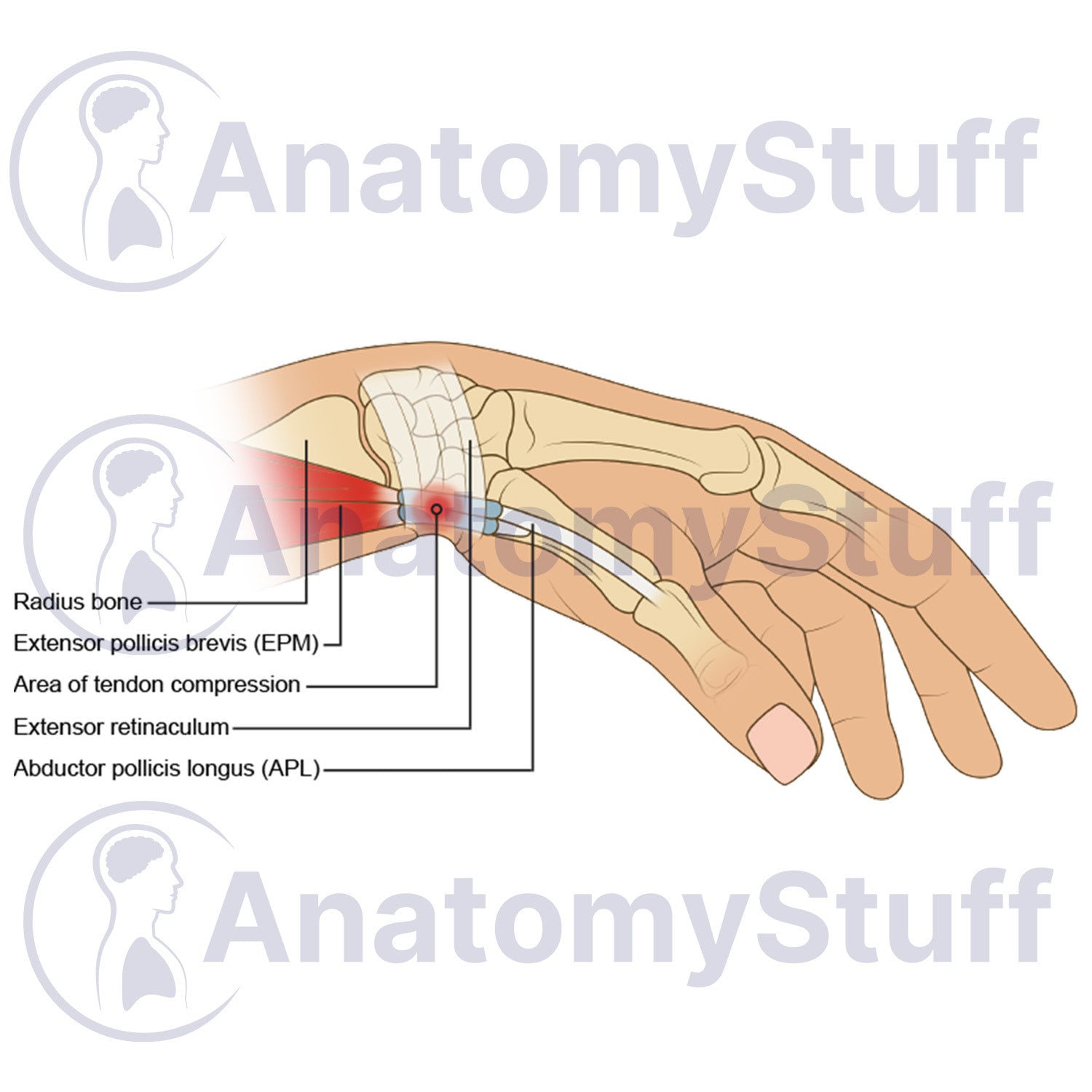
This licensable medical illustration depicts the anatomical structure of the human wrist and thumb, highlighting the area of tendon compression associated with De Quervain's tenosynovitis. Optimised for medical education, this professional stock vector asset demonstrates the precise relationship between the radius bone, extensor retinaculum, extensor pollicis brevis, and abductor pollicis longus tendons. It is ideal for healthcare educators, orthopaedic studies, clinical training courses, and physiotherapy practices looking for detailed upper limb anatomical graphics.
Product Specifications
- Format: PNG (Transparent background)
- Dimensions: 1100 x 800 px
- Resolution: 300 DPI
- Print Size: ~10 x 8 cm
- Colour Profile: RGB (Optimised for digital and print)
- File Size: ~280 KB
Licensing Information
Please select the licence that matches your intended use:
- Science Licence: Licence for academic purposes such as theses research publishing, and the scientific discourse.
- Education Licence: Licence for educational purposes, live teaching, presentations, handouts, and exam papers.
Commercial Use: Interested in using this for advertising, book publication, or other commercial purposes? Please Contact Us to discuss a Commercial Licence.
Please allow 1-2 working days for delivery of your image.
By purchasing, you agree to our Licensing Terms and Conditions.