






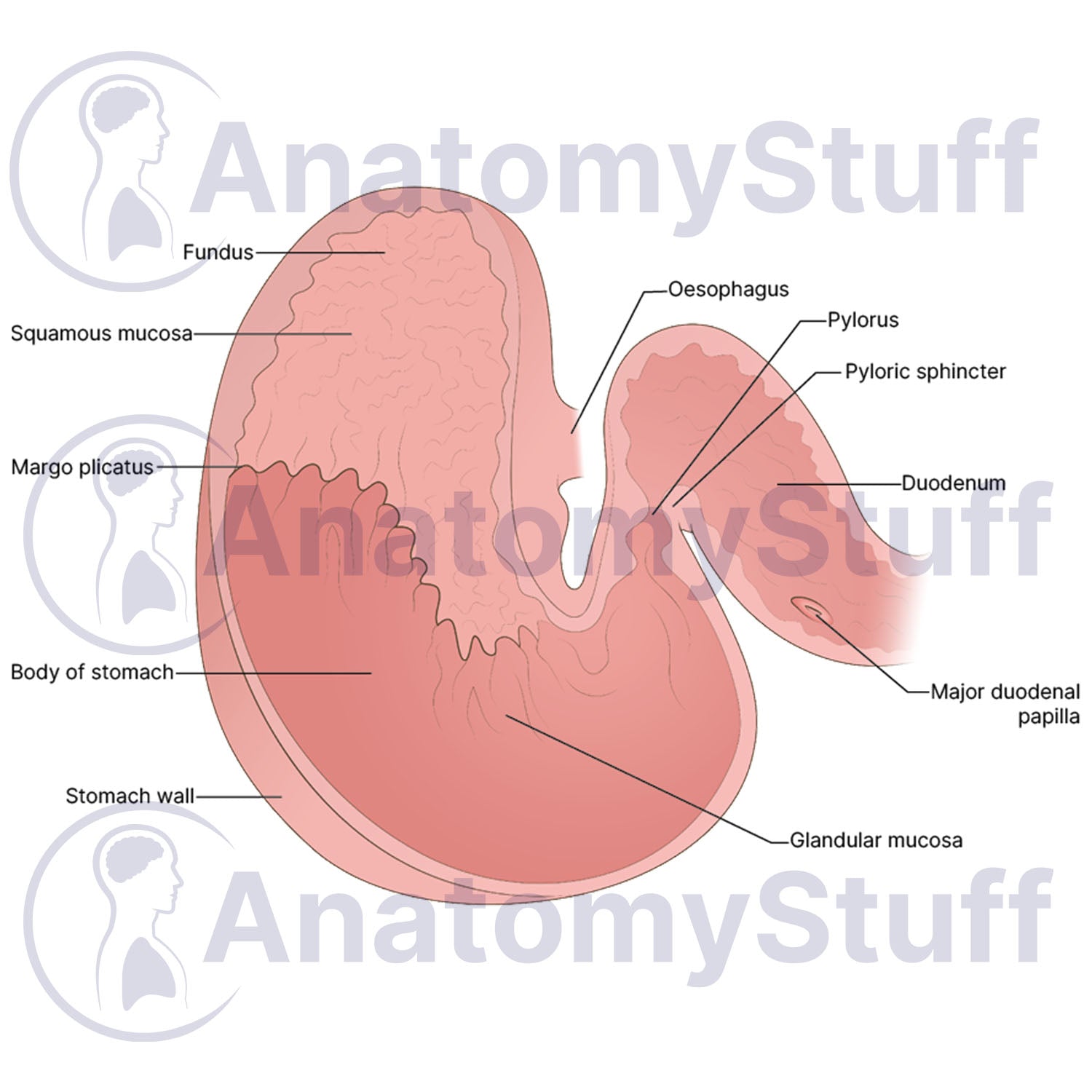
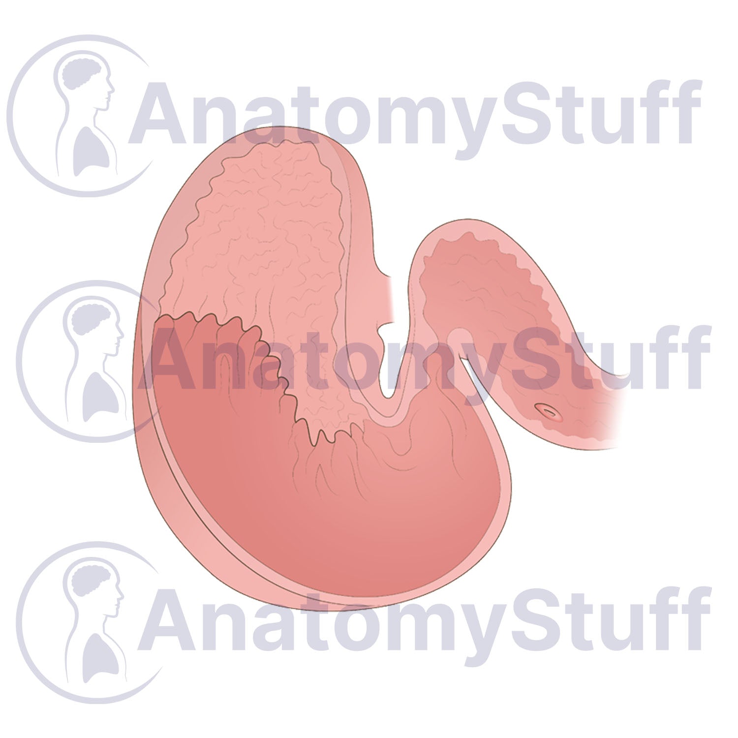


This scientific illustration of Equine Stomach Anatomy provides a clear, comprehensive view of the horse's internal gastric system. Illustrated by a professional medical illustrator, this horse internal stomach anatomy view is perfect for equine educators, veterinary students, researchers, and horse care professionals.
Anatomical Highlights
- Mucosal Division: Clearly demonstrates the distinct margo plicatus separating the non-glandular squamous mucosa from the glandular mucosa region.
- Gastric Structure: Highlights key internal regions including the fundus, the main body of the stomach, and the thick stomach wall layers.
- Inflow and Outflow Pathways: Details the junction from the oesophagus down through the pylorus, pyloric sphincter, and into the duodenum with the major duodenal papilla.
Choose Your Variant
- Fully Labelled: Ready for immediate use in presentations.
- Unlabelled (Blank): Perfect for interactive learning. This clean version is ideal for student examinations, "fill-in-the-blank" quizzes, or custom labelling for specialised research.
Product Specifications
- Format: PNG (Transparent background)
- Dimensions: 1181 x 945 px
- Resolution: 300 DPI
- Print Size: ~8 x 10 cm
- Colour Profile: RGB (Optimised for digital and print)
- File Size: ~300 KB (Blank) ~350 KB (Labelled)
Licensing Information
Please select the licence that matches your intended use:
- Science Licence: Licence for academic purposes such as theses research publishing, and the scientific discourse.
- Education Licence: Licence for educational purposes, live teaching, presentations, handouts, and exam papers.
Commercial Use: Interested in using this for advertising, book publication, or other commercial purposes? Please Contact Us to discuss a Commercial Licence.
Please allow 1-2 working days for delivery of your image.
By purchasing, you agree to our Licensing Terms and Conditions.


