


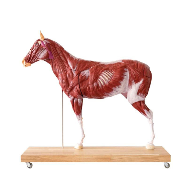
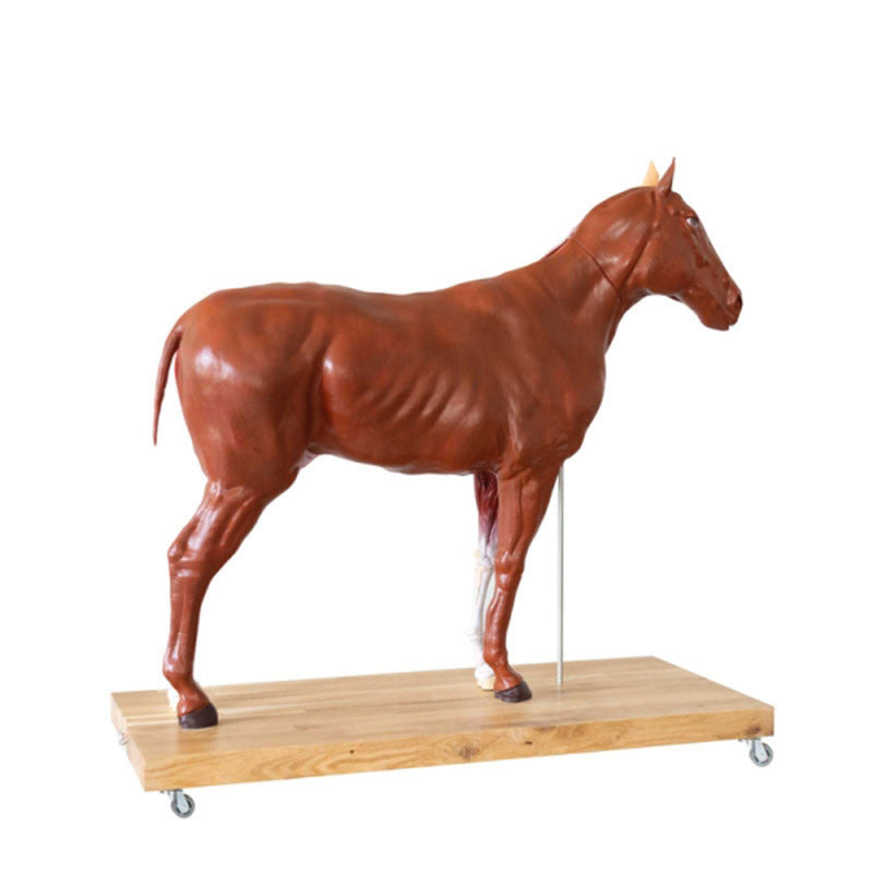
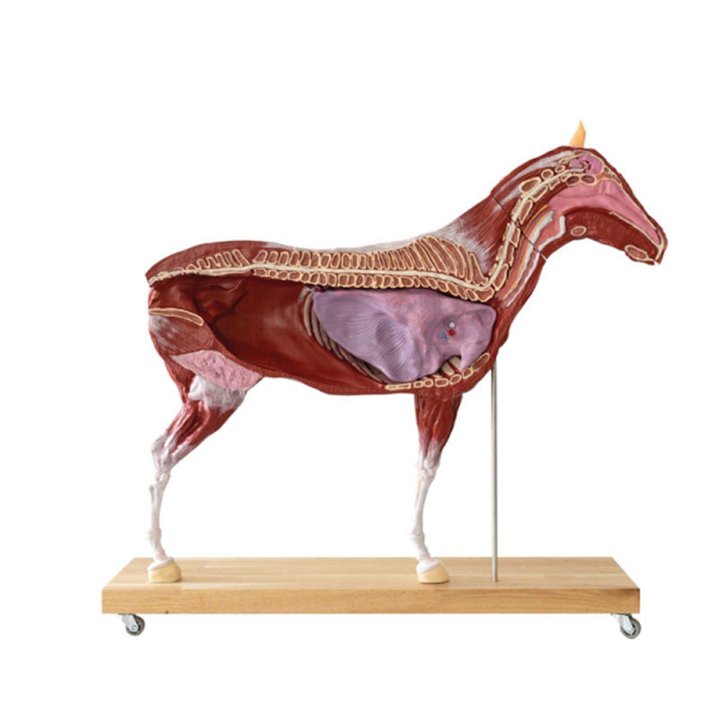
This unique 16 Part, 1/3 Life-Size Female Horse Model has been created for teaching and demonstrating internal and external anatomy to veterinary students. Created on a sturdy base with wheels this equine model can be split into two parts along the sagittal plane.
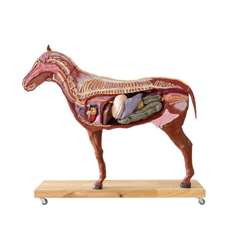
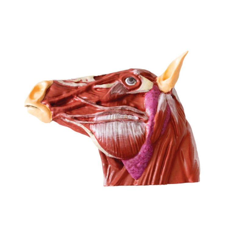
The right side of the sagittal plane shows the external skin of the animal, whilst the left side of the sagittal plane shows the body and limb muscles. When the two halves are divided the left side shows the chest and abdominal cavity, this cavity includes detachable organs (listed below). The right side of the horse model shows the main blood vessels, the horse's heart and the abdominal cavity with detachable organs. In addition to these features, half of the horse's head can be detached from the main model, allowing students to see the muscles.
This equine model can be separated into 16 individual parts.
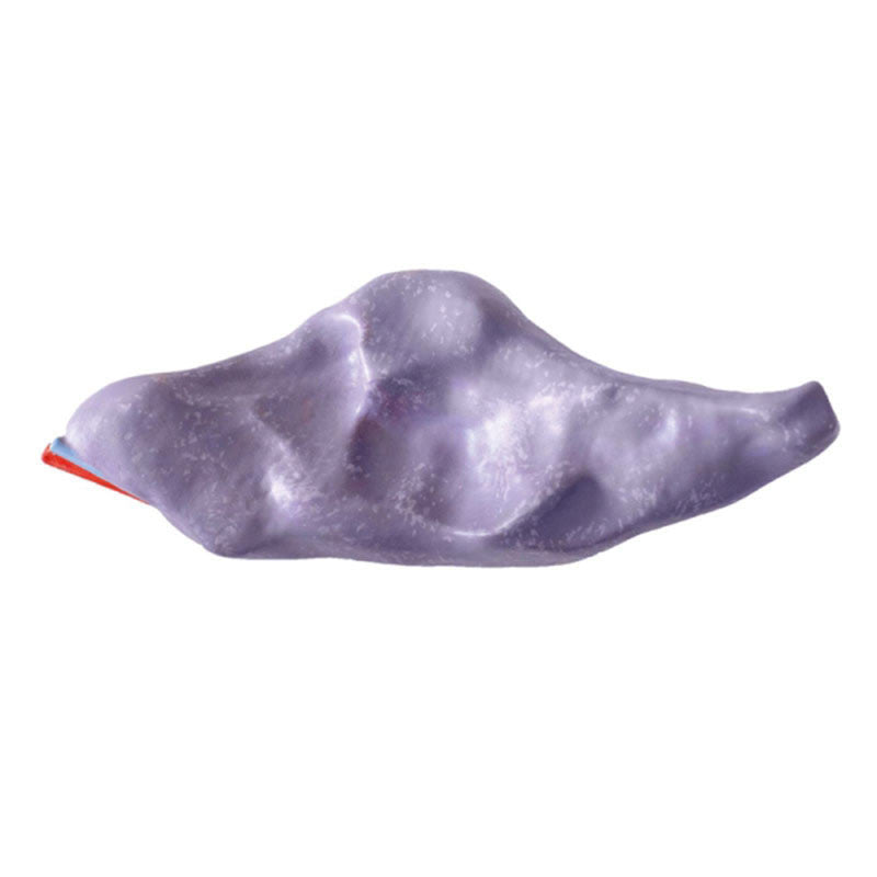
- Right side of the head - external
- Left side of the head - internal muscles
- Right side of the trunk - external
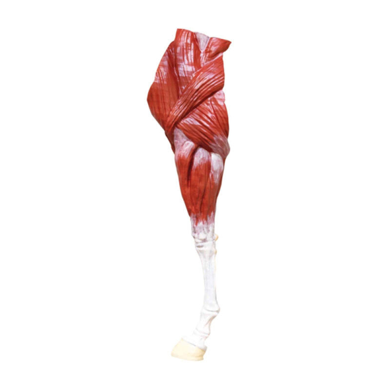
- Left side of the trunk - internal muscles
- Left front leg
- Right gluteal muscle
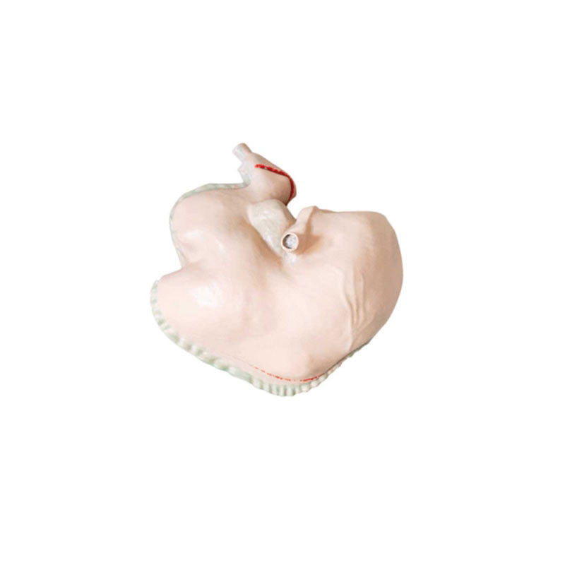
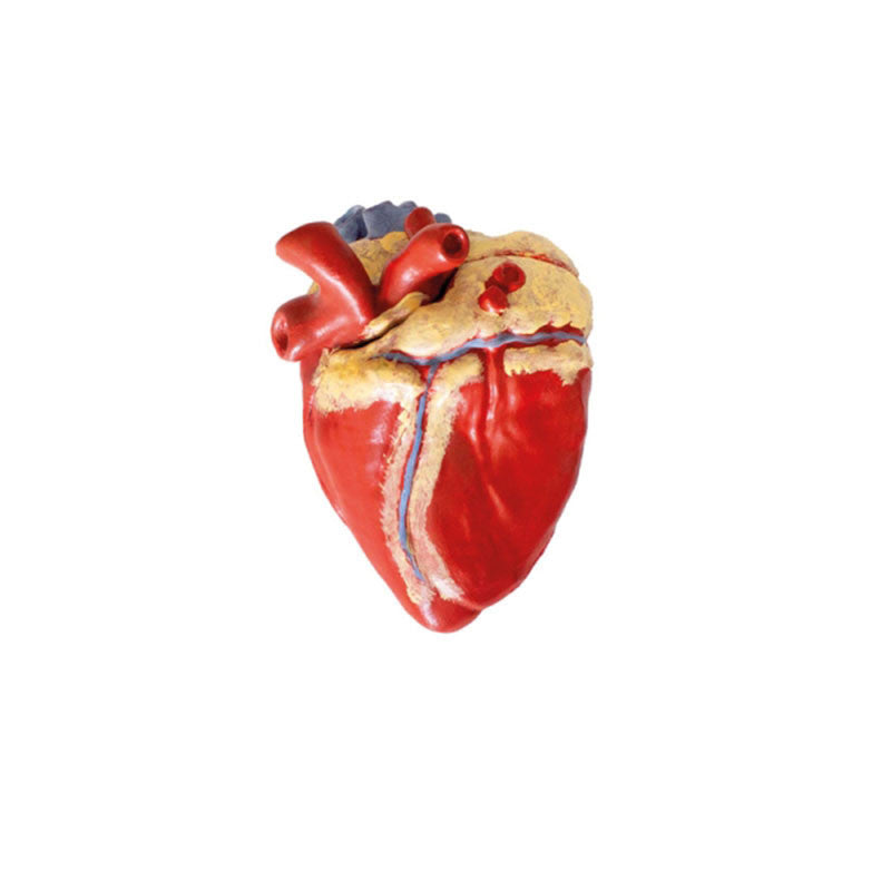
- Heart - 2 parts
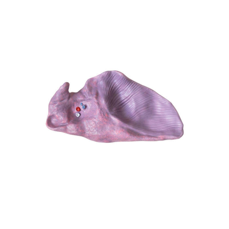
- Left lung
- Stomach - 2 parts
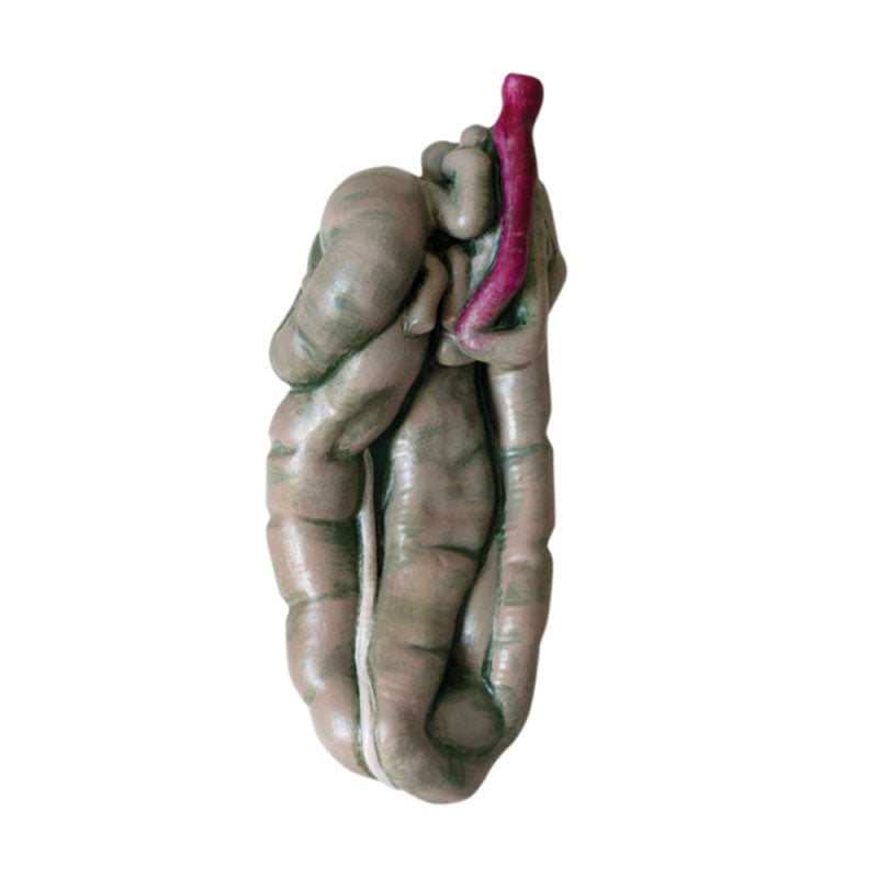
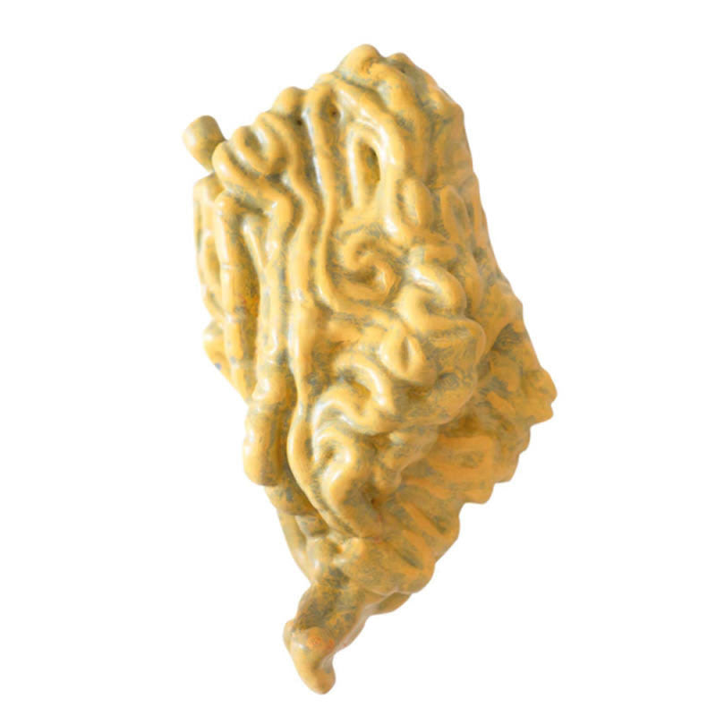
- Small intestine
- Large intestine
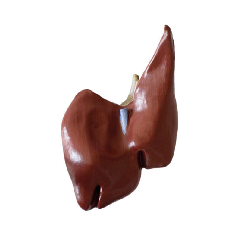
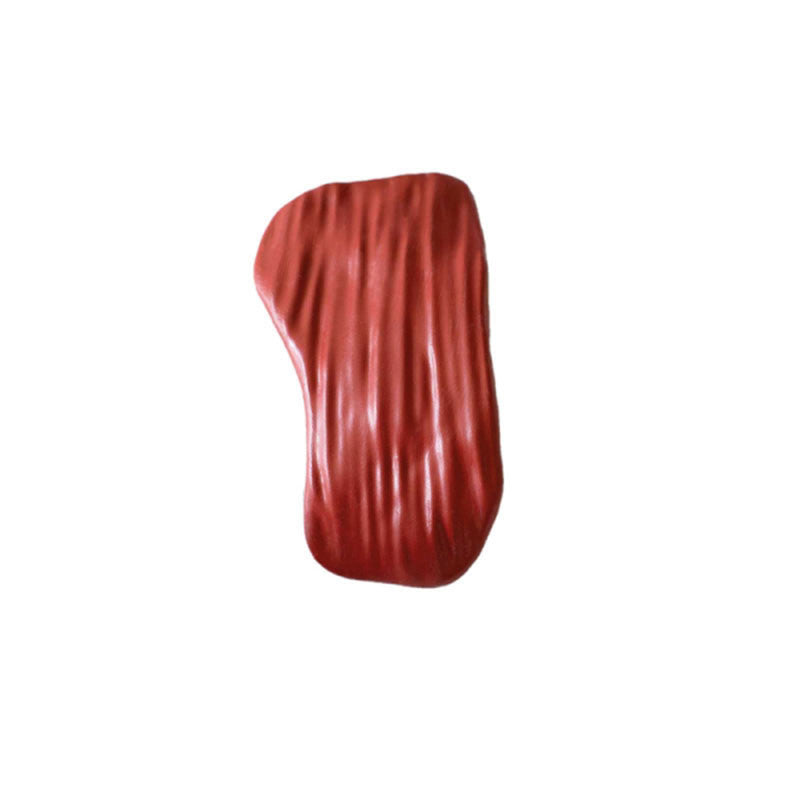
- Liver
- Left kidney
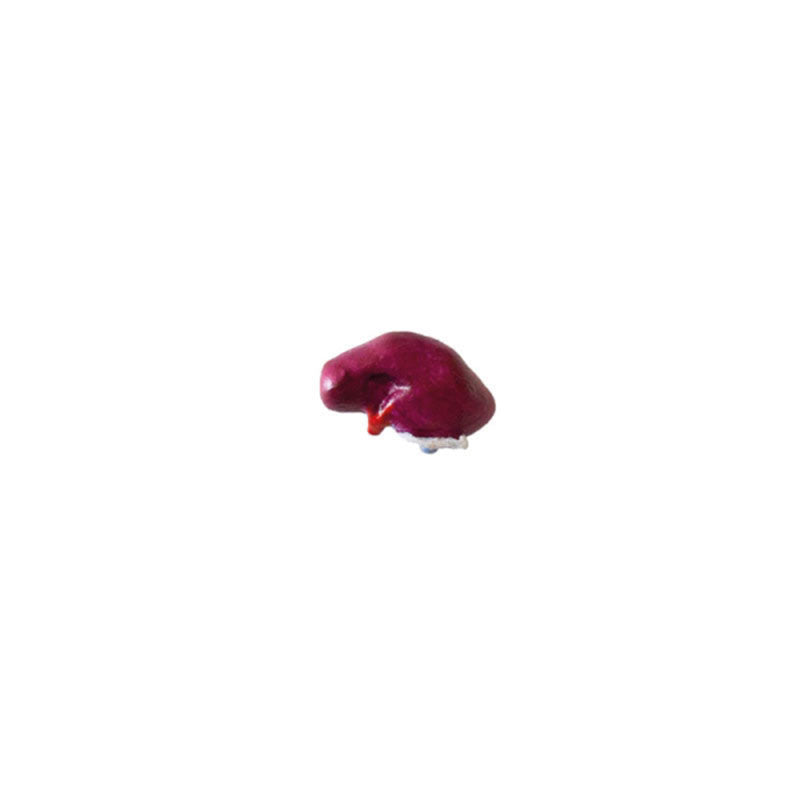
- Spleen
Our veterinary range also includes a Bovine Model, Pig Model and Sheep Model.