


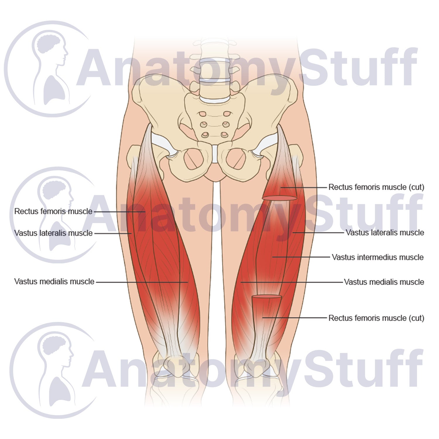
This quadriceps anatomy stock illustration provides a clear anterior view of the human thigh musculature. The diagram simplifies the visual mapping of the anterior compartment for sports science students, physiotherapy universities, and clinical lecturers. It offers an accurate tool for demonstrating both superficial and deep muscular layers of the lower limb during live teaching and assessment.
Anatomical Features
- Superficial musculature: Anterior view showing the intact rectus femoris, vastus lateralis, and vastus medialis muscles in relation to the pelvic girdle.
- Deep muscle dissection: Sectioned view displaying the cut rectus femoris to expose the underlying vastus intermedius muscle layer.
- Bony landmarks: Clear structural positioning showing muscle origins and pathways relative to the hip and femur bones.
Product Specifications
- Format: PNG (Transparent background)
- Dimensions: 1100 x 800 px
- Resolution: 300 DPI
- Print Size: ~10 x 8 cm
- Colour Profile: RGB (Optimised for digital and print)
- File Size: ~300 KB
Licensing Information
Please select the licence that matches your intended use:
- Science Licence: Licence for academic purposes such as theses research publishing, and the scientific discourse.
- Education Licence: Licence for educational purposes, live teaching, presentations, handouts, and exam papers.
Commercial Use: Interested in using this for advertising, book publication, or other commercial purposes? Please Contact Us to discuss a Commercial Licence.
Please allow 1-2 working days for delivery of your image.
By purchasing, you agree to our Licensing Terms and Conditions.